Refractive
Spring 2024
by Ellen Stodola
Editorial Co-Director
Creating a true accommodating lens has been a goal for some time in ophthalmology. Several ophthalmologists discussed some of the products currently in development in this space and what makes them unique.
Juvene lens by LensGen
Source: Sumit “Sam” Garg, MD, and LensGen
This lens is in two parts, said Sumit “Sam” Garg, MD, with a capsule-filling haptic that has an optic in it and also a receptacle for the action part of the lens, which is a fluid lens.
Basically, you put that into the capsular bag, and that maintains the capsular volume, Dr. Garg said. Inside that, in a two-part insertion, you place the fluid lens into the eye and then tab it into place. “There are three tabs, and you pop it into place and that’s what supports the lens inside the eye,” he said. The anterior membrane is deformable, and with accommodative effort, it becomes steeper, which gives you the ability to get this range of vision—distance, intermediate, near.
The Juvene lens is a silicone and non-diffractive lens. “Some of the limitations we have with the diffractive optics and presbyopia correction go away because it’s non-diffractive, so you’re not as concerned about other pathologies,” he said. “But on the flip side, you get monofocal-like optics with multifocal-like range, and the conversation becomes easier.”
Right now, when people talk about lenses, they usually talk about the great range achieved but rarely marry that with the visual quality performance, Dr. Garg said. “They’ll say, ‘I get great quality,’ but don’t talk about the range. This will allow us to do both at the same time, and because of the proposed mechanism of action, I think that’s really intuitive for patients.”
Dr. Garg said the Juvene lens takes a bit of patient adaptation. “I think it takes a little while for the eye to get used to, at least what we’ve seen in initial trials,” he said. “It’s not necessarily like you put it in and the next day you have full range. Most people see improvement in near and intermediate over the first month or so, but really after 3 months is when it starts to kick in.”
The Juvene lens is not currently in an active trial but the company is ramping up for a Phase 1 FDA trial. All the data published so far has been in exploratory study outside U.S., Dr. Garg said, so it’s a different patient population but favorable data. He added that data on the lens has been presented at the ASCRS Annual Meeting and has been published in the Journal of Cataract & Refractive Surgery. “We recently presented 3-year data at ASCRS [and] showed maintenance of magnitude of effect, so whatever accommodation you had at year 1, you saw at year 3,” he said, adding that results were the same in respect to contrast sensitivity.
What is seen with this lens is binocular summation, he said, so if you have it in both eyes, those patients as a whole did better than patients who just had it in one eye. “I think that’s true of most of these lenses. This is not a monovision effect. This is a distance-corrected actual effect of the lens,” Dr. Garg said.
The incision for the Juvene lens is around 3–3.2 mm, Dr. Garg said, adding that “the only real learning curve with the surgery is getting used to the two-part insertion and tabbing it in the eye.” In terms of safety, he said endothelial cell counts are similar to traditional surgery, and there hasn’t been any major inflammation. You don’t get significant PCO because the bag stays open, he said. “This is not just true of the Juvene; it’s true for any of these lenses that keep the bag open.”
So far, the results have been promising with the published data, he said.
JelliSee IOL by JelliSee Ophthalmics
John Vukich, MD, has been working with the trial of the JelliSee IOL, which he said is a lens that is designed to mimic the natural lens of a child. In its resting state the lens will, by design, be focused for near. One of the key features of the design is that it does not rely on whole bag compliance, capsular contraction; it does not rely on the posterior capsule; it is activated by a change in the diameter of the capsular bag complex. “The lens is fundamentally different, as it’s mechanism of action is actually potentiated by fibrosis of the lens capsule.”
JelliSee, he said, has undergone an initial clinical trial, and there is data from both primates and humans. These studies were conducted in El Salvador, where Dr. Vukich participated as the primary surgeon, having done all the cases to date.
Dr. Vukich has been involved with the development, surgical technique, postop care, and data associated with the JelliSee IOL, and said that there is currently 1-year data on 10 patients. “We are able to demonstrate reliably up to 7 D of accommodative change,” he said.
This lens, he added, can be used for anyone who has a need for cataract surgery, and, at some point, refractive lens exchange may become an indication as well. “One of the fundamental design features of the lens is that the change in the shape occurs on the anterior surface of the implant, and the activating force is applied by the capsular ring complex anterior to the equator of the lens,” he said. “The lens achieves an enhanced level of translation of the applied force by moving the point of contact with the lens anterior to the equator. It results in a more dynamic anterior surface change. The lens design optimizes the translation of mechanical force from the ciliary body creating a much more efficient ability to change the shape of the anterior surface of the lens.”
Multiple studies have shown that the ciliary muscle remains strong and functions throughout life and is able to exert, on average, 0.08 Newtons of radial force. The lens is designed to use this available force by creating eight points of contact within the peripheral capsular bag. The posterior capsule and capsulotomies do not contribute to the mechanism of action. “As the capsular bag fibroses with time, it potentiates the translation of the ciliary body movement into the capsular ring diameter, and the lens becomes more efficient. Rather than a concern about capsular bag fibrosis leading to a disadvantage of the mechanism of action, it’s actually the exact opposite,” he said. “It improves the mechanism of action, and we are seeing that both with the longer-term primate studies as well as in our human studies.”
The lens is designed so that 114 microns of total diameter change is enough to activate the entire potential of the lens, he added. That is what is needed for the full dynamic 7 diopter range of the lens, he said. “Even patients who can generate only half the average amount of force will still be able to achieve 4.5–5 D of accommodative changes. It’s a very robust system that doesn’t require an average response; even a suboptimal response will still provide a significant diopter change.”
Dr. Vukich said that all patients who have been treated with this lens have been done with only three different base powers. The large dynamic range means that we can target the patient for hyperopia and the patient will then naturally focus for distance. The best-corrected visual acuity in all patients was 20/20 or better, he said, and near is a natural function that occurs in the first day or two postoperatively and becomes more robust at 1 month.
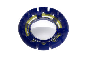
OmniVu modular shape-changing lens system by Atia Vision

a fixed-power front optic and fluid-filled,
shape-changing base lens
Source: George Waring IV, MD, and Atia Vision
The OmniVu modular shape changing lens system is comprised of a fixed-power front optic and fluid-filled, shape-changing base lens, making it somewhat unique in the marketplace and developmental pipeline, according to George Waring IV, MD.
“The goal is to treat cataract and address the effects of presbyopia by harnessing the natural lens accommodating mechanism of the eye, and the way this works is that there is a fixed-power front optic hydrophobic acrylic, and it has docking tabs that are around the periphery of the optic,” he said. “Then, there’s a shape-changing, fluid-filled base.” The front fixed-power optic docks into that, and it’s a modular system that allows for customization in the future, he said. This lens is designed to restore a full range of vision and minimize visual disturbances that can be seen with other presbyopia-correcting solutions, Dr. Waring said. When the body attempts to read, you have the ciliary muscles exert a force on the capsular bag, and that translates to a shape-changing base that results in the base optic thickening in curvature, and that’s what increases the power for reading, he added.
The OmniVu lens has been evaluated in pilot studies, and Dr. Waring noted that there is 1-year data for two sites. It’s currently designed for patients with under 1.5 D of astigmatism and is being looked at for opportunities for a toric platform in the future, he said. The power range is currently 12–28 D, coming in half diopter increments. Efforts are underway to assemble the U.S. IDE trial.
Dr. Waring noted that there are some nuances to the surgical procedure, but overall, this is the same fundamental principles of phacoemulsification and cataract surgery. Since it’s a two-piece modular lens system, it’s done in two steps utilizing a 3.5-mm incision and then an intended 5.5-mm manual capsulotomy. You do want to pay particular attention to be careful with incision size and also capsulotomy diameter, he said.
Because of the nature of the optics, this has an excellent visual quality, contrast sensitivity, and dysphotopsia profile, Dr. Waring said. “In addition, it seems there are some lesser understood benefits of capsular filling technology in terms of effective lens position and reduction of posterior capsular opacification, and that’s very exciting for the future,” he said.
Results so far have been positive. Dr. Waring said he presented 6-month outcomes at the 2023 ASCRS Annual Meeting, showcasing that 95% of eyes were within a half diopter of plano at 6 months.
Ocumetics accommodating lens by Ocumetics Technology Corp.
The Ocumetics accommodating lens is a shape-changing lens that was developed by Dr. Garth Webb, beginning in 2007, said R. Doyle Stulting, MD, PhD. The lens is implanted into the capsular bag. “When the ciliary body contracts, tension on the lens zonules is reduced, and the refractive elements of the Ocumetics lens increase its refractive power through a patented process involving a small, air-filled space,” Dr. Stulting said. It is constructed of solid silicone and does not contain liquid silicone or any potentially toxic substance, he said, and the change in refractive power is almost instantaneous.
Dr. Stulting said that the simple design of the lens mimics the natural crystalline lens. “It is contained completely within the capsular bag, does not impinge on the ciliary processes, does not contain any liquids that might escape, does not involve any electronics, and rapidly changes its power in response to ciliary body contraction/relaxation,” he said.
Any patient undergoing cataract surgery would be a candidate for implantation of the Ocumetics lens, Dr. Stulting said. “Current implants either do not permit clear near vision (monofocal IOLs), optically split incoming rays of light between near- and distance-focusing elements of the lens (multifocal lenses), or optically expand the range of focus,” he said. “None of these products provides sharp near and distance vision without compromise.” All ophthalmologists are aware of visual aberrations attributable to these lenses, some of which lead to lens explantation. At some point in the future, the Ocumetics lens may even be used to treat presbyopia in individuals who have not yet developed cataracts, he added.
Dr. Stulting said the lens requires minimal to no adjustment from patients, since the Ocumetics lens mimics closely the action of the natural crystalline lens.
The lens has undergone pre-clinical testing, but first-in-human trials have not yet begun. These are planned for Q2 2024, Dr. Stulting said.
About the physicians
Sumit “Sam” Garg, MD
EyeWorld Chief Medical Editor
Vice Chair of Clinical Ophthalmology
Medical Director
Director of Technology
Professor – Cataract, Corneal & Refractive Surgery
Gavin Herbert Eye Institute
University of California, Irvine
Irvine, California
R. Doyle Stulting, MD, PhD
Chief Medical Officer
Ocumetics Technology Corp.
Founder, Stulting Research Center at Woolfson Eye Institute
Professor of Ophthalmology, Emeritus
Emory University
Loudon, Tennessee
John Vukich, MD
Founding Partner Central American Ophthalmic Research Consultants
Summit Eye Care
Madison, Wisconsin
George Waring IV, MD
Waring Vision Institute
Mt. Pleasant, South Carolina
Relevant disclosures
Garg: LensGen
Stulting: Ocumetics
Vukich: JelliSee Ophthalmics
Waring: Atia Vision
Contact
Garg: gargs@hs.uci.edu
Stulting: dstulting@icloud.com
Vukich: javukich@gmail.com
Waring: georgewaringiv@gmail.com