Cataract
Spring 2024
by Ellen Stodola
Editorial Co-Director
Foreign body sensation (FBS) is a common issue that may be associated with a wide range of conditions and may present with different signs and symptoms. Two physicians discussed this problem, including when they’re most likely to see it and how to diagnose it.
“It’s difficult to guess percentage, but I think virtually all patients will experience some form of irritation at some point in their life including FBS, and in my experience, there is a specific subgroup, roughly 10%, who have a severe form that is recalcitrant to treatment,” said D. Brian Kim, MD.

Source: D. Brian Kim, MD
The persistence of FBS implies chronicity, he said, which may include dry eye syndrome, blepharitis, or allergic conjunctivitis. Anatomical comorbidities involving the eyelid such as floppy eyelid syndrome, conjunctivochalasis, epithelial basement membrane dystrophy, and recurrent corneal erosions can exacerbate the problem as well.
Rony Sayegh, MD, said that persistent FBS is when an individual experiences the sensation that there is something gritty, scratchy, or uncomfortable in their eye, even when no actual foreign object is present. The prevalence of persistent FBS can vary depending on the underlying causes and the population being studied, he said, adding that it’s a relatively common complaint that ophthalmologists encounter. He also noted dry eye as one of the most common causes, affecting a significant portion of the population.

Source: Rony Sayegh, MD
How much does cataract surgery contribute?
Dr. Sayegh said that FBS of varying severity, persisting for 3 or more months after cataract surgery, seems to occur in about 10–15% of patients. Although the mechanism is not fully understood, there are a number of possible reasons for this postoperative FBS, he said.
Local edema over the incision from surgical trauma is one possible factor, and microcystic edema, or even frank bullae, can be seen on exam. Hypertonic saline solution can be helpful in these cases as the edema is often transient.
Persistent inflammation is another cause that is not well understood. Patients with a history of dry eye, diabetes, underlying connective tissue disease, or certain nutritional alterations may be at a higher risk.
Damage to corneal nerves at the incision site is another potential cause. Severing corneal nerves can lead to alterations in the functional tear unit with decreased tear production, reflex tearing, and decreased release of neuroprotective agents, all resulting in dry eye and damage to the ocular surface that can be perceived as FBS. Furthermore, aberrant regeneration of the severed nerves and possibly neuroma formation can result in persistent FBS.
Dr. Kim also discussed how cataract surgery can exacerbate the problem. “First, a whole host of medicated eye drops are given preoperatively, intraoperatively, and postoperatively, and these drops contain preservatives, which are toxic to the ocular surface. Betadine can also irritate the eye,” he said. “Second, the very act of surgery severs corneal nerves, which can reduce corneal sensation. Moreover, cutting the cornea can damage the epithelial basement membrane adhesions and result in a recurrent corneal erosion.” He added that typically, recurrent corneal erosion is associated with accidental injury, but it’s important not to forget that surgery is also a form of trauma to the cornea, particularly clear corneal phaco. “The most important precaution is to identify comorbid conditions and counsel patients during the preoperative discussion, so they are made aware and begin treatment before surgery.”
Source: Rony Sayegh, MD
Presentation and common complaints
In some cases, patients will explicitly say, “I never had FBS until after you did my cataract surgery,” Dr. Kim said. This is a challenge because even if you improve the patient’s visual acuity, they may dwell on the FBS, which they will reiterate only began after you did surgery. Some of this can be subclinical dry eye or blepharitis manifested from surgery, while others can have an underlying epithelial basement membrane dystrophy or corneal erosion.
“A good history including a good description of symptoms, when they began, how they’ve progressed, and any factors that seem to worsen or alleviate the discomfort, underlying health issues, and environmental exposures can be very helpful,” Dr. Sayegh said. “In addition, resolution of the FBS with instillation of a drop of anesthetic in the office is often helpful in narrowing the differential diagnosis. Slit lamp examination, which includes eyelid eversion as well as use of vital dyes, can help make the diagnosis in the great majority of patients.” Dr. Sayegh said that more specific diagnostics such as tear osmolarity, MMP-9, meibomian gland imaging, and confocal microscopy can also be helpful.
When diagnosing FBS, Dr. Kim said it’s important to first treat the common conditions, namely dry eye and blepharitis. However, even after aggressive treatment, patients may not improve, and the patient and physician can be left perplexed and frustrated, he said. “It’s important to consider that these cases could actually be a form of recurrent corneal erosion not visible on slit lamp examination.”
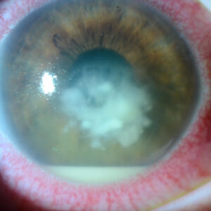
Typically, Dr. Kim said that recurrent corneal erosions are diagnosed by identifying irregular or negative staining with fluorescein dye or looking for irregularities on corneal topography. However, he noted that identification relies on a high index of suspicion since there is no good definitive diagnostic tool. A dried Weck-Cel sponge has been used to touch the corneal surface to identify areas of loose epithelium, but the dry sponge can stick to the cornea and induce an abrasion. “As a result, I developed the Kim Corneal Sweeper [Corza Medical], which is a handheld instrument with a straight handle and fusiform shape tip with rounded edges,” he said. “The smooth texture enables safe use on the corneal surface. Fluorescein dye is instilled, and the cornea is examined under the slit lamp with cobalt blue light. The sweeper is held like a pencil and pressed tangentially against the cornea with steady indentation pressure along the entire corneal surface. The tear film enables it to glide over the cornea atraumatically. When an area of loose epithelium is encountered, it ripples up, which creates a visible fold easily seen due to the thin profile and enhanced by the fluorescein dye. Typically, the area of loose epithelium is isolated and well-demarcated. We published a study using the corneal sweeper to detect these hard-to-find corneal erosions and discovered that more than 80% of these patients experienced some improvement or resolution of symptoms after treatment.1”
See “Corneal sweep test for recurrent corneal erosion” from the June 2022 issue of EyeWorld for more details on the technique.

Source: Rony Sayegh, MD
Management process
The treatment for FBS depends on the underlying cause. Since foreign body sensation can be due to various factors, Dr. Sayegh suggested some general approaches and treatments:
1. Identify and treat the underlying condition. Remove any foreign bodies and exposed concretions, optimize the ocular surface. Treat conditions appropriately with antibiotics, antihistamines, and/or steroids. Hypertonic saline can help with corneal edema and epithelial basement membrane dystrophy. Use lid hygiene for meibomian gland dysfunction and blepharitis. Response to therapy can sometimes help determine the main underlying cause of the FBS, and optimizing that line of treatment, whether it is using plugs in patients who find artificial tears helpful or cyclosporine in patients who find steroids helpful, can increase the odds of success.
2. Placing a drop of proparacaine in the eye and asking if it helps relieve the FBS can help diagnose corneal nerve dysfunction. Warm compresses not only help with blepharitis and meibomian gland dysfunction but seem to help reduce nerve pain as well. Autologous serum tears remain the most effective treatment for the neuropathic form of FBS and have been shown to help regenerate corneal nerves on confocal microscopy. Scleral lenses can also be helpful in some cases.
3. Avoid rubbing. Rubbing the eyes can exacerbate irritation and FBS. Dr. Sayegh has used wristbands that vibrate when patients attempt to touch their eyes (designed for trichotillomania) with some success.
Article Sidebar
Persistent foreign body sensation in the eye can be caused by a variety of conditions and factors, Dr. Sayegh said. Some of the most common ones include:
Dry eye syndrome: Dry eye is a broad term that includes any condition in which tears are impacted, in quantity, quality, composition, or distribution on the ocular surface. Blepharitis, meibomian gland dysfunction, and conjunctivochalasis are very common conditions that are part of the dry eye syndrome, and a gritty or sandy feeling in the eyes is commonly reported by patients with these conditions.
Conjunctivitis: This includes allergies, which are very common, as well as viral conjunctivitis. Patients often report itching, irritation, redness, and the sensation of a foreign body in the eye. Everting the eyelids can be helpful in the diagnosis of these conditions to look for follicles and papillae, Dr. Sayegh said.
Contact lens issues: FBS can be a symptom of a poor fit or contact lens spoilage or related to a complication of contact lens wear, such as a corneal abrasion or ulcer. It is also important to ask if wearing the contact lens helps with the FBS, which can occur in some patients, such as those with limbal stem cell deficiency.
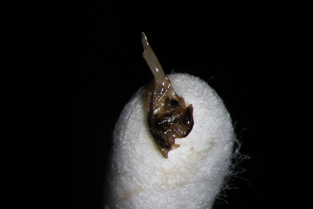
Foreign body: Sometimes a small foreign object can actually get stuck in the eye or under the eyelid, leading to FBS. “We have seen contact lenses retained for decades. One should not forget conjunctival concretions are common and can sometime break through the surface and cause FBS,” he said.
Environmental factors: Wind, smoke, dust, and other environmental factors can cause foreign body sensation. “Some of my patients who work long hours outdoors or at the grill have benefited from protective goggles,” he said.
Eye strain: Prolonged periods of reading or using digital screens can cause eye strain, leading to discomfort which may include FBS.
Corneal erosions: These are also a common cause of FBS and include epithelial basement membrane dystrophy and recurrent erosion syndrome. It is important to look carefully for patterns of negative staining that is indicative of these conditions, he said. Other dystrophies are often easier to diagnose based on corneal appearance. Certain systemic drugs such as novel cancer treatments can also cause corneal changes and FBS.
About the physicians
D. Brian Kim, MD
Professional Eye Associates
Dalton, Georgia
Rony Sayegh, MD
Cole Eye Institute
Cleveland Clinic
Cleveland, Ohio
Reference
- Kim ME, Kim DB. Implementation of the corneal sweep test in the diagnosis of recurrent corneal erosion: a 2-year retrospective study. Cornea. 2022;41:1248–1254.
Relevant disclosures
Kim: None
Sayegh: None
Contact
Kim: docdbk100@gmail.com
Sayegh: rrs109@case.edu