Glaucoma: Complicated cases
September 2023
by Ellen Stodola
Editorial Co-Director
According to glaucoma specialists Mary Qiu, MD, Aakriti Shukla, MD, and Catherine Sun, MD, neovascular glaucoma (NVG) is an aggressive, secondary glaucoma that develops in the setting of retinal ischemia. Dr. Sun discussed signs and strategies to manage this condition, and ophthalmology residents Alexis Kassotis, MD, and Jessie Wang, MD, also shared their thoughts on this topic.
Signs and characteristics
The most common etiologies that lead to NVG are proliferative diabetic retinopathy (PDR), retinal vein occlusion (RVO), and ocular ischemic syndrome (OIS),1 Dr. Sun said. “Many patients are diagnosed with NVG when they present with an acute rise in IOP, severe eye pain, and/or decreased vision,” she said. “These symptoms are often due to neovascularization of the angle (NVA), which obstructs aqueous outflow through the normal drainage pathway of the eye and leads to elevation in IOP and severe pain.”
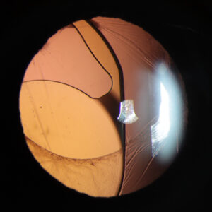
Aside from the normal eye exam features, it is important to look for neovascularization of the iris (NVI) and NVA on slit lamp exam before dilation and by performing gonioscopy, Dr. Sun said. Gonioscopy can tell you if there is NVA or synechial angle closure. Other signs of NVG include conjunctival hyperemia or corneal edema if an acute rise in IOP occurs, although this may not be present if the IOP elevation is insidious. Hyphema or microhyphema often occur due to bleeding of fragile anterior segment vessels, and ectropion uveae or corectopia may be seen if synechiae exist,2 she added. A dilated exam should be performed after gonioscopy to determine if there are other complications such as vitreous hemorrhage, retinal detachment, or macular edema, and to help determine the etiology of the condition.
Early stage disease is characterized by anterior segment neovascularization with normal IOP. NVI usually starts at the pupillary border with vessel growth in a disorganized fashion. In some cases, NVA can be present without NVI.2 As a fibrovascular membrane continues to grow over the trabecular meshwork, IOP can rise, Dr. Kassotis said. In the earliest stages, there are no peripheral anterior synechiae, but as the disease progresses, peripheral anterior synechiae can develop. “The presence of synechiae leads to partial or complete angle closure, while their absence denotes open angle disease,” she said. “At present, this terminology exists with or without the presence of glaucomatous optic neuropathy (i.e., once there is glaucomatous optic neuropathy, it is termed a ‘glaucoma’).”
Dr. Sun, along with Dr. Qiu and Dr. Shukla, advocate for a consensus panel to standardize the nomenclature for NVG.
Management
Dr. Sun, Dr. Qiu, and Dr. Shukla said that the management of NVG includes: 1) controlling the IOP, and 2) addressing the underlying disease responsible for the neovascularization.
“To control the IOP, topical and oral medications are used initially. If medical therapy is not enough, which is usually the case, or if patients need to be placed on oral carbonic anhydrase inhibitors, laser or surgical treatments should be offered,” she said. “These traditionally include cyclophotocoagulation (CPC), aqueous shunts, or trabeculectomy.”
Dr. Sun said that valved aqueous shunts are generally preferred as the definitive treatment given high rates of failure with trabeculectomy and immediate pressure lowering with valved compared to non-valved aqueous shunts. CPC is usually reserved for patients with poor visual potential and may have a temporary effect. “In rare cases, I will perform CPC in an emergent situation in the clinic or emergency room if medications are not effective and we are unable to take the patient to the operating room urgently,” she said. “If the effect of CPC is not enough, patients will generally get a subsequent aqueous shunt.”
Dr. Qiu said that in her practice pattern, in eyes with total synechial angle closure at the time of NVG presentation and active anterior segment neovascularization, she will perform gentle CPC in the OR setting with judicious steroids, regardless of visual potential. “I agree if the effect of CPC is not enough to control the IOP, patients will get an aqueous shunt later,” she said. “I prefer to use a non-valved aqueous shunt with a 3-0 Prolene ripcord to prevent early and late hypotony.” There are many different ways to approach surgical management of eyes with NVG, Dr. Qiu said, and we need more data to learn which way results in the best patient outcomes.
Dr. Sun stressed that addressing the underlying disease requires working with retinal colleagues. “These patients generally need anti-VEGF injections and/or panretinal photocoagulation (PRP), depending on the underlying condition,” she said. “They may need systemic work-up and control of their systemic disease, too.” For NVG, immediate anti-VEGF injection at the time of presentation is often helpful to save any portion of the angle that is still open because anti-VEGF injections work faster than PRP to regress neovascularization.
Dr. Wang said that patients with NVG require a multidisciplinary treatment approach. Patients must be followed closely from the retina perspective to treat the underlying retinal pathology causing the ischemia and neovascularization. This includes a combination of intravitreal anti-VEGF injections to rapidly regress the neovascularization, as well as PRP to address the ischemia more permanently, she said.
Simultaneously, patients require prompt and close follow-up with a glaucoma specialist to normalize the IOP. “How this is achieved depends largely on the angle anatomy at presentation. In patients with completely open or even partially open angles, the IOP responds fairly well to medical therapy. This means that topical IOP-lowering medications and/or oral carbonic anhydrase inhibitors can be used first, providing some leeway and time for anti-VEGF agents to regress the active neovascularization so that surgery can be performed in a more controlled context with a lower bleeding risk.”
Since the angle is still fully or partially open, Dr. Qiu and Dr. Wang suggest that performing an angle surgery to restore the conventional outflow pathway can be considered. There are treatment protocols being piloted that may allow this to be successful, Dr. Qiu said. In patients with completely closed angles, the IOP doesn’t decrease appreciably with medical therapy or correlate with the regression of active neovascularization following anti-VEGF therapy. “Patients in this category will require surgical intervention, but in an eye with active anterior segment neovascularization, risk of bleeding- associated complications remain high,” she said. One strategy that can rapidly lower the IOP while also allowing the neovascularization to regress before performing incisional surgery is to implement a staged approach consisting of prompt gentle cyclophotocoagulation to rapidly normalize the IOP and prevent ongoing glaucomatous optic neuropathy, followed by implantation of a tube shunt at a later date, if needed, once the eye is quiescent.
How does MIGS fit into the treatment paradigm?
There was a case report in a patient with a partially open angle who successfully underwent gonioscopy-assisted transluminal trabeculotomy (GATT) to restore aqueous outflow from her conventional outflow pathway,3 but more research is needed in this area, Dr. Qiu said. “The rationale for possible angle surgery in the earlier stages of NVG is that the trabecular meshwork is at least partially open without extensive peripheral anterior synechiae. Since fibrovascular proliferation and tissue contraction has not yet completely blocked the angle, the angle can still be potentially salvaged by cutting through the fibrovascular membrane that is blocking the trabecular meshwork and restoring aqueous outflow. However, if there are active anterior segment neovessels, angle surgery is not advised because the rate of bleeding-associated complications is very high. We recommend anti-VEGF injections to regress the neovessels first, and angle surgery should only be considered in eyes that have quiescent disease with no active anterior segment neovessels.”
She added that the underlying disease process needs to be aggressively treated by the retina team to prevent the neovessels from returning, which can be challenging. These patients need to understand that if GATT fails, they will need a subsequent, more definitive surgery, such as an aqueous shunt to control the IOP.
MIGS may be an option for patients with neovascular glaucoma with completely or partially open angles, Dr. Wang said. Rather than “giving up” on the conventional outflow pathway when the angle is still functioning, glaucoma specialists can utilize MIGS to restore the conventional outflow pathway. Dr. Wang noted GATT as a potential option because it requires access to only a small portion of the angle in order to access Schlemm’s canal but can then open up the angle across the entire 360 degrees. However, many patients with NVG may be on chronic blood thinners, which increases the risk of bleeding-associated complications after GATT, and Dr. Qiu does not offer GATT in patients who cannot hold anticoagulation.
Dr. Qiu and Dr. Wang added that MIGS cannot be performed in eyes with complete synechial closure of the angle. “In this challenging cohort of patients, medications likewise often do not adequately lower the IOP,” Dr. Wang said. “In those patients with synechial closure and IOP too high for the health of the optic nerve, prompt laser procedures such as CPC and incisional surgery such as tube shunt implantation are standard of care. Finally, during the treatment of neovascular glaucoma, follow-up with the retina service is crucial to control the underlying ischemic retinal disease.” She noted that failure to do so often leads to recurrence of neovascularization and new formation of peripheral anterior synechiae, which would lead to failure of the MIGS that has been performed.
Anti-VEGF and PRP for patients with neovascular glaucoma
Dr. Sun said that anti-VEGF injections are given intravitreally and can lead to rapid regression of neovascularization. These include regression of NVI and NVA. “However, if synechiae angle closure has already developed, anti-VEGF injections may be less effective at normalizing IOP. They will cause regression of NVI or NVA but the synechiae angle closure will remain,” she said.
Dr. Kassotis said that in normal aqueous humor, VEGF is absent, while high levels of VEGF accumulate in the aqueous of those with ischemic anterior segment conditions, such as NVG. VEGF induces angiogenesis in response to the relative hypoxia, leading to iris and angle neovascularization and increased IOP. Given VEGF’s integral role in NVG pathogenesis, VEGF inhibition has emerged as an important modality in NVG treatment.4,5
Anti-VEGF leads to rapid reduction in anterior segment neovascularization, Dr. Kassotis said, adding that anti-VEGF agents can be administered as intravitreal or intracameral injections.4 While angiography demonstrates that anti-VEGF significantly decreases leakage of the aberrant vessels, vessel complexes do not regress completely, unlike in panretinal photocoagulation.
Management of NVG depends on visual potential and patient comfort, Dr. Kassotis said. “Aggressive medical management with topical and oral IOP-lowering agents is helpful acutely, often as a segue to more definitive management,” she said. While anti-VEGF is helpful in the short term for open angle NVG, PRP has been used in ischemic disease for decades. It is efficacious both in the early and more advanced stages of NVG (including closed angle NVG), thus is considered a mainstay of treatment despite the advances of anti-VEGF. PRP reduces aqueous VEGF levels more than anti-VEGF and structurally reduces the number of anterior segment neovessels.6
Optimizing outcomes
Depending on the underlying etiology for retinal ischemia, additional systemic treatment may be needed, Dr. Sun said, adding that PDR is the most common cause of NVG. “For these patients with diabetes mellitus, their blood glucose and blood pressure should be controlled,” she said. “I recommend that these patients see an endocrinologist if they do not have one.”
Dr. Sun stressed that prevention is the most important way to stop vision loss from NVG. “This includes education to patients who have conditions that make them at high risk for NVG, such as diabetic retinopathy and retinal vein occlusion,” she said. “For non-glaucoma providers who care for these patients, incorporating gonioscopy into routine practice in patients who are high risk is important for the earliest diagnosis. If early neovessels can be detected before the angle has synechially closed, these patients may have a chance of preserving their natural aqueous outflow pathway and not need glaucoma surgery.”
There are a number of other challenges to optimal care for NVG patients once they develop NVG that she, Dr. Qiu, and Dr. Shukla discussed in an editorial in Ophthalmology Glaucoma.7 “We felt that ‘multidisciplinary discussions in ophthalmology are needed around the following topics: (1) standardizing the definition and staging of NVG; (2) detecting anterior segment neovascularization earlier; (3) increasing evidence-based research to improve outcomes; (4) determining the optimal multidisciplinary treatment approach; and (5) increasing patient adherence to treatment.’”
About the physicians
Alexis Kassotis
PGY-2 Ophthalmology Resident
Edward Harkness Eye Institute at Columbia University Medical Center
New York, New York
Mary Qiu, MD
Glaucoma Specialist
Department of Ophthalmology and Visual Science at University of Chicago
Chicago, Illinois
Aakriti Garg Shukla, MD
Glaucoma Specialist
Edward Harkness Eye Institute at Columbia University Medical Center
New York, New York
Catherine Sun, MD
Glaucoma Specialist
Department of Ophthalmology at University of California, San Francisco
San Francisco, California
Jessie Wang, MD
PGY-4 Ophthalmology Resident
Department of Ophthalmology and Visual Science at University of Chicago
Chicago, Illinois
References
- Vancea PP, Abu-Taleb A. Current trends in neovascular glaucoma treatment. Rev Med Chir Soc Med Nat Iasi. 2005;109:264–268.
- Aboobakar IF, Lin MM. Clinical diagnosis of neovascular glaucoma in the ophthalmology office. In: Qiu, M. (eds) Neovascular Glaucoma. Essentials in Ophthalmology. 2022.
- Kanter J, et al. Gonioscopy-assisted transluminal trabeculotomy in neovascular glaucoma: Salvaging the conventional outflow pathway. Am J Ophthalmol Case Rep. 2022;28:101668.
- Viruni N, Cai CX. Anti-vascular endothelial growth factor for neovascular glaucoma. In: Qiu, M. (eds) Neovascular Glaucoma. Essentials in Ophthalmology. 2022.
- Grisanti S, et al. Intracameral bevacizumab for iris rubeosis. Am J Ophthalmol. 2006;142:158–160.
- Olmos LC, et al. Long-term outcomes of neovascular glaucoma treated with and without intravitreal bevacizumab. Eye (Lond). 2016;30:463–472.
- Qiu M, et al. Improving outcomes in neovascular glaucoma. Ophthalmol Glaucoma. 2022;5:125–127.
Relevant disclosures
Kassotis: None
Qiu: None
Shukla: None
Sun: None
Wang: None
Contact
Kassotis: ak4129@cumc.columbia.edu
Qiu: mary.qiu@gmail.com
Shukla: ag2965@cumc.columbia.edu
Sun: Catherine.Sun@ucsf.edu
Wang: jessie.wang@uchicagomedicine.org