Refractive
Spring 2024
by Liz Hillman
Editorial Co-Director

Source: William Trattler, MD
Among the important indications for use of the EVO ICL (STAAR Surgical)—age, amount of myopia, astigmatism correction, etc.—there is one parameter that some surgeons say could be causing confusion.
“Yes, I do think there can be some confusion,” Audrey Rostov, MD, said.
“Yes, absolutely. It’s confusing,” William Trattler, MD, said.
“100% agree … there is significant confusion,” Scott D. Barnes, MD, said.
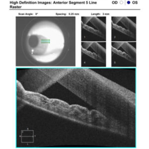
The FDA’s indication specifies that the EVO ICL should be used in patients “with an anterior chamber depth of 3.0 mm or greater.” It further defines this as “when measured from the corneal endothelium to the anterior surface of the crystalline lens.”
“There is confusion because nobody agrees on what the term ‘anterior chamber depth’ really means. Does it include the epithelium or is it measured just from the endothelium? Depending on which textbook and which literature report you look at, it may say anterior chamber depth is measured from the endothelium,” Dr. Barnes said, adding later that some instruments report ACD from epithelium to anterior capsule and others have the ability to subtract the cornea and report the measurement from the endothelium or both. “Yes, absolutely there is confusion on it, and some instruments cannot separate that out and they just record ACD. Some instruments separate it out and report ACD endo vs. ACD epi (as with Orbscan); others report ACD internal vs. external, as with Pentacam where “ext” includes the corneal thickness and “int” is from the endothelium. Some others will also display AQD or AD, meaning ‘aqueous depth,’ which means the ACD without the corneal thickness. … You have to know your machines and what they’re reporting.”
Dr. Rostov said similar. In terms of sizing for the ICL, she said, “it’s important to know that you should be measuring from the endothelium, not the epithelium. You want to make sure about this because otherwise you could choose the wrong size lens or you may not even have enough anterior chamber depth to permit use of the ICL.”
Dr. Rostov said she uses Pentacam (Oculus) to measure anterior chamber depth from the endothelium as well as white to white. If a surgeon is using biometry for these measurements, like the IOLMaster (Carl Zeiss Meditec), improper sizing could occur. Dr. Rostov has found that white to white measurements with biometers is generally larger.


Source: Scott D. Barnes, MD
With Pentacam, Dr. Rostov said she can set it to measure from endothelium (internal). If you were to use a device that measures from the epithelium (external cornea), you need to subtract out the pachymetry of the cornea, she said.
“The reason this is important is you want to make sure you get right sizing of ICL,” Dr. Rostov said. “If you put in ICL and you don’t have enough anterior chamber depth, you could get narrowing of anterior chamber. The vault of the ICL could be too high. In a shallow anterior chamber, you have the potential for angle closure glaucoma. Make sure you have enough AC depth. Strict on-label use of the ICL is a 3-mm anterior chamber depth as measured from the endothelium.”
Blake Williamson, MD, said his practice actually found out that it had been inputting data from the ACD exterior (measuring from the epithelium) instead of interior. Thankfully, he added, it didn’t have a clinical impact.
“When you have a technology that’s as amazing as the EVO ICL, frankly, it makes up for a lot of minor things that you might be doing wrong because patients are seeing so well and because it’s so forgiving,” he said. “But even though it’s so forgiving and patients are going to be happy to ecstatic, it doesn’t forgive not being really careful with how you’re entering the data. So it’s important to have this conversation about how we enter the data.”
If you are using a device that measures from the epithelium, Dr. Barnes said to simply subtract the corneal thickness and use that number in your nomogram. He said the STAAR OCOS software displays a warning reminder to “please enter the true ACD” and defines this as measured from the endothelium” and to subtract the corneal thickness if your instrument only measures ACD from the epithelium.


Source: Scott D. Barnes, MD
Dr. Trattler said he has been using the LASSO formula that takes data from the IOLMaster 700 and the MS-39 anterior segment OCT (Costruzione Strumenti Oftalmici, CSO), which is input into an excel spreadsheet. The formula predicts the expected vaults for each different size ICL. With this formula, he said he has felt comfortable implanting a number of ICLs in patients who were below the 3.0, on-label ACD indication in the U.S. Dr. Trattler explained that the on-label ACD was the limit set for inclusion in the clinical trials to help make sure patients ended up with a normal vault. “Now that we have more data both within the U.S. and internationally, as well as more analysis of EVO ICL outcomes, we can use calculators to select ICLs that will have a normal vault, even if the ACD is below 3.0,” he said.
Erik Mertens, MD, said internationally an ACD (endothelium to anterior capsule) of 2.8 mm for myopia and 3.0 for hyperopia is acceptable. He, however, finds “you can go lower than the ACD recommended by STAAR if the angle is wide open.” “Understanding the true ACD means investigating the morphology and width of the ciliary sulcus and the ciliary body,” Dr. Mertens continued.
Dr. Mertens said in an email to EyeWorld he will consider ICL for patients who have smaller ACD measurements after a thorough discussion and, again, if the angles are wide open.
“With anterior segment OCT measurements and UBM (VuMAX, Sonomed Escalon) to check the ciliary body and sulcus in combination with new calculation methods based on AI, we’re getting very good predictions in how the vault will be after surgery,” said Dr. Mertens who uses the LASSO formula to calculate his IOL sizes.1 “Lower vault, even down to 100 µm, is not considered an issue with ICLs with a central hole, and most of experts would only intervene if the ICL was touching the crystalline lens; otherwise, with good aqueous flow over the natural lens, monitoring is preferred.”
Overall, Dr. Barnes said it’s important for surgeons using EVO to remember to track their data and their outcomes, adjusting, if necessary, when they see trends, similar to how they would adjust their surgeon factor and laser nomogram after tracking outcomes for cataract or laser refractive surgery. In the end however, he said that there is such forgiveness with EVO that even with vaults higher or shallower than we’d like, it may be like +0.25 D or –0.25 D after cataract surgery. “It’s different with EVO; high and low vaults with the legacy ICL were associated with clinical issues (IOP issues and/or lens opacities) more frequently than we are seeing with the EVO ICL,” Dr. Barnes said.
Article Sidebar
Sizing insights
Dr. Rostov said she tends to size down with EVO by one with non-toric versions, while with toric she’ll go with the size recommended by the STAAR calculator.
“If a toric rotates, that’s more problematic,” she said.
She has found cataract formation with EVO to be “quite low,” and stability and placement working well when downsizing non-toric versions. “Not everyone does this,” she said. “I like to be conservative and would rather undersize than oversize. I have had a couple of torics where I undersized and they did rotate.”
Sizing down, when possible, she said reduces the risk of having too high of a vault and the potential for angle closure, she said.
Dr. Williamson said his practice also routinely sizes down. “Just remember low vault, low problems. Big vault, big problems.”
His other pearl was to be consistent in the devices you’re using to obtain measurements among patients. “If you’re consistent and it’s working for you, stick with that,” he said.
For more details and perspectives on EVO sizing, read “Taking a closer look at ICL sizing and vault concerns” on page 72.
About the physicians
Scott D. Barnes, MD
Chief Medical Officer, STAAR Surgical
Colonel (retired), U.S. Army
Womack Army Medical Center
Fort Bragg, North Carolina
New York, New York
Erik Mertens, MD, FEBOphth
Medical Director
Medipolis
Antwerp, Belgium
Audrey Rostov, MD
Cornea, Cataract, and Refractive Surgeon
Seattle, Washington
William Trattler, MD
Director of Cornea
Center for Excellence in Eye Care
Miami, Florida
Blake Williamson, MD
Williamson Eye Center
Baton Rouge, Louisiana
Reference
- Rocamora L, et al. Postoperative vault prediction for phakic implantable collamer lens surgery: LASSO formulas. J Cataract Refract Surg. Feb 1 2023;49(2):126–132.
Relevant disclosures
Barnes: STAAR Surgical
Mertens: STAAR Surgical
Rostov: STAAR Surgical, Carl Zeiss Meditec, Alcon, Bausch + Lomb
Trattler: STAAR Surgical, Oculus, Carl Zeiss Meditec, CSO
Williamson: STAAR Surgical
Contact
Barnes: sbarnes@staar.com
Mertens: E.Mertens@medipolis.be
Rostov: audreyrostov@gmail.com
Trattler: wtrattler@gmail.com
Williamson: blakewilliamson@weceye.com