Ophthalmology News
January 2008
by Maxine Lipner
EyeWorld Senior Contributing Editor
Three different staining patterns help practitioners to determine dry eye severity
The pattern of lissamine green staining can help point the way toward a dry eye diagnosis, according to James P. McCulley, M.D., professor and chairman, department of ophthalmology, University of Texas Southwestern Medical School, Dallas. In the July 2007 issue of Eye and Contact Lenses, Dr. McCulley described three distinct patterns to be on the lookout for with lissamine green.
Lissamine and other vital stains have an important role in pegging cases of dry eye, Dr. McCulley believes. “Dry eye in its objectively established phase has dry spots on the surface, and the lissamine green will stain where the devitalized dry cells are,” he said.
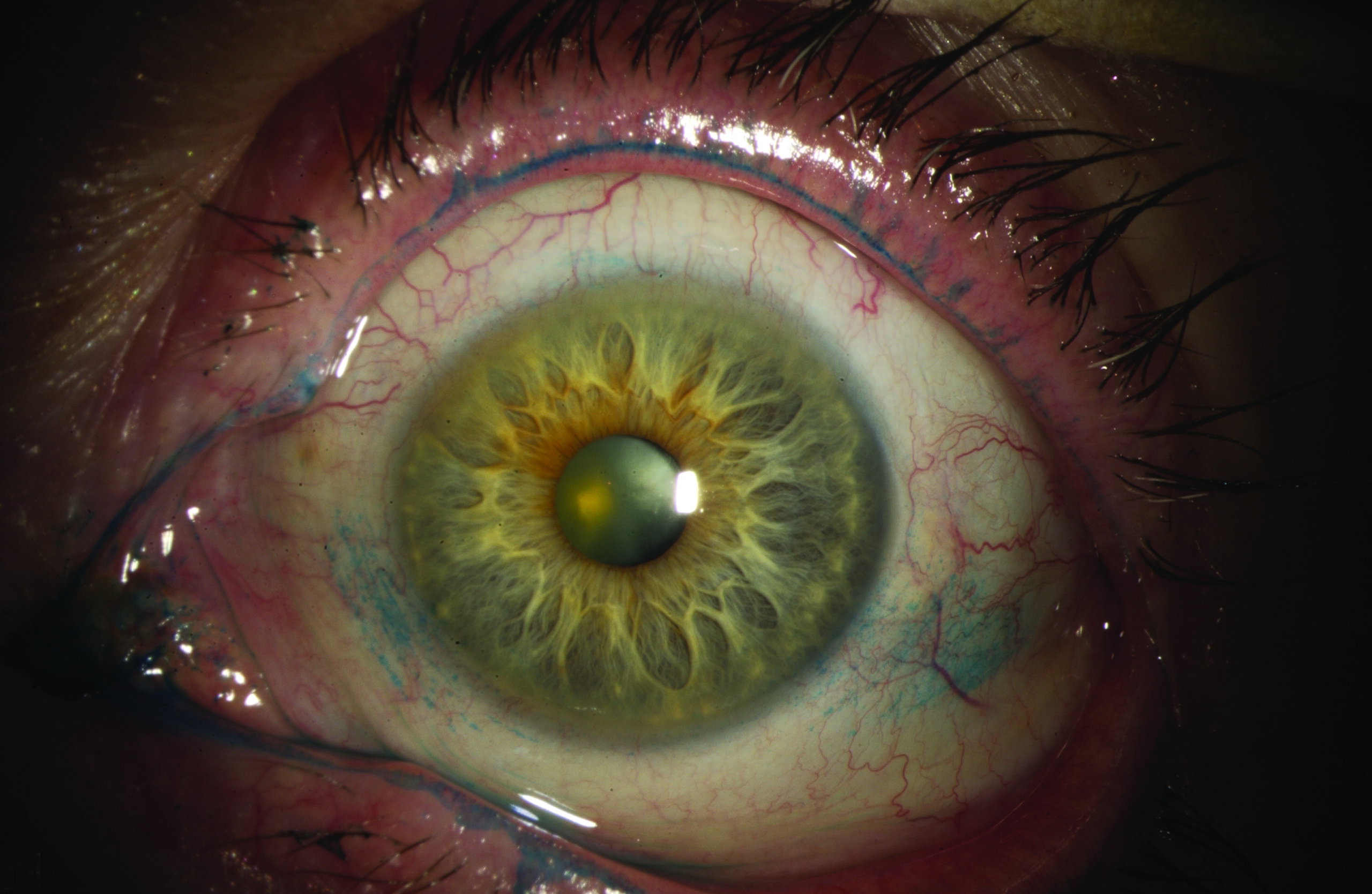
Source: James P. McCulley, M.D.
Patterns come into view
It was after noticing that specific staining patterns among his office patients seemed to emerge depending upon the severity of the dry eye case that Dr. McCulley decided that a closer look at this was warranted. “I had become increasingly impressed over my years as an ophthalmologist that if there was interpalpebral conjunctival staining of the conjunctiva nasally, that that could be non-specific, or could be the earliest clinical evidence of a dry eye condition,” Dr. McCulley said. “However, this was relatively non-specific, whereas, if there was both nasal and temporal interpalpebral staining that that was pretty definitely dry eye in the absence of some other obvious reason for the temporal staining.” The latter also seemed to mark a more advanced stage of the disease. Corneal staining meant that the disease had progressed even further. Dr. McCulley and fellow investigators examined staining patterns in 22 dry eye patients and 11 control subjects. “We did the study to look at the aqueous tear volume and to assess whether in fact there was objective evidence for this clinical impression of the (progressive) staging of an aqueous deficient dry eye,” he said. “Indeed we found evidence for that.”
Investigators found that as the vital staining score increased, there was a progressive decrease in the Shirmer’s test result. Also, they determined that there was an increase in meibomian gland dropout among dry eye patients when compared to control subjects.
The research in this study ultimately bolstered Dr. McCulley’s observations among patients in his practice, indicating that it had been on target. “Clinical observation led to a clinical laboratory assessment that bore out our hypothesis, that nasal staining alone was the mildest form of aqueous deficient dry eye,” he said. “Also that nasal and temporal staining indicated intermediate, and nasal, temporal, plus cornea were the most significant.”
Comparing stains
Lissamine green is, of course, not the only stain in town. Another vital stain available is rose bengal. Dr. McCulley finds that these both have excellent staining ability and sees them as preferable to fluorescein. “Both lissamine green and rose bengal will allow us to see the dry spots on the conjunctiva as well as the cornea,” he said. “Whereas fluorescein will really only effectively allow us to see the staining on the cornea.” As a result, if practitioners rely on fluorescein staining, they are in effect delaying the dry eye diagnosis. “We won’t have objective evidence of ocular surface drying and dry spots if we only use fluorescein until the dry eye is much more advanced to severe,” Dr. McCulley said. He sees lissamine green and rose bengal as more or less interchangeable and both as being superior to fluorescein. The same patterns that Dr. McCulley spotted with the lissamine green also mark the spot with rose bengal staining. The one difference between lissamine green and rose bengal is the stinging sensation that patients often experience with the latter. “Lissamine tends to be better tolerated by the patient; there is less irritation,” Dr. McCulley said.
While these study results should translate well to the ophthalmic office, making it easier for practitioners to pick out the real dry eye patients, there is one practical consideration to keep in mind: vital dyes such as lissamine green and rose bengal must be specially ordered. “There’s not a commercially available drop of either lissamine green or rose bengal,” Dr. McCulley said. “But they can be obtained from formulating pharmacies.” There are, however, commercially available strips available. “The problem is getting enough dye off the strips onto the surface of the eye,” Dr. McCulley said. “So I much favor a formulating pharmacy’s creation of a drop that gets enough dye in to effectively stain the surface, if there is surface staining.” While he is not a big fan of the strips, Dr. McCulley does see these as better than nothing if a practitioner does not wish to approach a formulating pharmacy.
Overall, Dr. McCulley believes that the study has important clinical implications in helping practitioners to ferret out dry eye patients sooner. “Ophthalmologists can avoid a lot of unexpected problems with keratorefractive patients by insuring that if the patient has a mild dry eye before keratorefractive surgery is done that they’re aware of this and that they start dry eye therapy pre-operatively,” he said. “Otherwise, the keratorefractive surgery is almost certainly going to tip those mild dry eye patients over into moderate to severe dry eye patients post-operatively.”
Editor’s note
Dr. McCulley has no financial interests related to his comments.
Contact Information
Please contact EyeWorld editorial staff if you would like send correspondence to Dr. McCulley.