Cataract: Digital Momentum
Summer 2026
by Liz Hillman
Editorial Co-Director
Until recently, visualization during cataract surgery relied primarily on an analog surgical microscope, an instrument whose core optical design has remained largely unchanged for nearly a century, outside of incremental advances in optics and illumination.
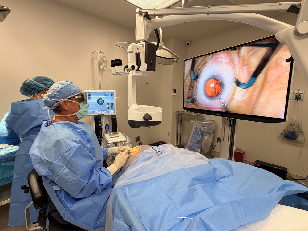
But digital visualization technologies that have already been on the market for several years are pushing cataract surgery toward increased precision, enhanced safety, and personalized care. These technologies not only benefit patient outcomes, but there are also advantages for the surgeon, including increased efficiency, better ergonomics, teaching and training opportunities, and more.
“Over the course of my career in ophthalmology, the transformation of intraoperative visualization technologies has been remarkable,” said Uday Devgan, MD, FACS. “When I began practicing, surgical visualization was largely dependent on conventional binocular operating microscopes with limited digital enhancement. Today, we’re witnessing a shift toward digital, immersive, and augmented visualization systems that have redefined how we approach anterior segment surgery.”
Source: Robert Weinstock, MD
Surgical visualization revolution
Dr. Devgan, Rosa Braga-Mele, MD, MEd, FRCSC, and Robert Weinstock, MD, shared their thoughts on and experience with digital operating technologies that have hit the market in the last decade—both wearable headset and heads-up display systems.
“The ergonomic benefits are notable—reduced neck and back strain, improved posture, and decreased physical fatigue. In addition, enhanced depth of field, digital contrast adjustments, and real-time filters improve the visualization of fine anatomical structures, particularly in complex cataract cases,” Dr. Devgan said.
Dr. Braga-Mele has experience with the NGENUITY 3D Visualization System (Alcon). While this system was originally developed for retina surgeons, Dr. Braga-Mele said it’s proved itself useful among cataract surgeons as well.
“It has a 3D TV screen visualization capability, and the newest software upgrades have improved light optimization, the ability to choose different color backgrounds to improve your depth of focus,” she said.
Dr. Braga-Mele spoke about the SeeLuma system (Bausch + Lomb), which she said offers digital binoculars or heads-up visualization on a screen, but its microscope arm is designed with a C-shape to allow for better screen positioning.
Dr. Weinstock has experience with several systems and said he performed the first cases in 2007 with the early prototype developed by TrueVision 3D Surgical. Since then, the technology has been refined, and the company has collaborated with other ophthalmic companies who have gone on to incorporate TrueVision technology or to create their own systems. TrueVision was acquired by Alcon and is now embedded in the NGENUITY 3D Visualization System.
In addition to the heads-up nature of these technologies, they often feature enhanced contrast and light optimization, data overlays, and workflow integration.
- Alcon NGENUITY 3D Visualization System: A 3D, heads-up system with the surgical field projected on a monitor
- Bausch + Lomb SeeLuma: A 3D, heads-up system projecting onto 4K monitors or for use with digital binoculars
- Beyeonics One: A wearable headset system for 3D surgical visualization
- Haag-Streit METIS 3D Digital Heads-Up Module: A 3D, heads-up system that attaches to the METIS 900 microscope for surgical visualization
- Leica Microsystems Proveo 8x: A 3D, heads-up system with the surgical field projected on a monitor
- Zeiss ARTEVO: A 3D, heads-up system with the surgical field projected on a monitor
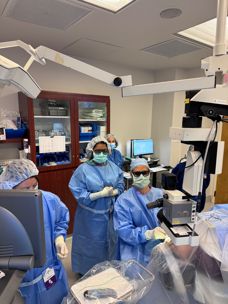
Dr. Braga-Mele said operating based on a heads-up display while wearing 3D glasses is much more ergonomic. “Your shoulders can be back, your neck can be more aligned because you’re not trying to compensate for bed height, and you’re not looking through binoculars,” she said. “It’s also nice because nurses in the OR and your assistant can be watching the screen in 3D while wearing the glasses as well.
“I have a very tall resident who has to stoop down, and if I’m assisting a tall resident, I almost have to stand up for the procedure to see through a regular microscope,” she said. “With heads-up technology, I can be sitting anywhere and watching at the same time.”
Improvements to operating microscopy through 3D, heads-up displays and headsets was sorely needed by surgeons. “In ophthalmology, musculoskeletal disorders are very prevalent … and it’s because of the way we sit at the slit lamp and at the microscope,” Dr. Braga-Mele said. “There are ergonomic chairs being developed that will marry with these ergonomic microscopes that we now have. To me, it’s exciting because it will maybe increase the longevity of the ophthalmologist, but it will definitely increase our quality of life.”
From a surgical and patient safety standpoint, Dr. Weinstock said 3D technologies have helped with use of high magnification for parts of the procedure such as capsule polishing or finding the edge of a capsulorhexis tear.
“When you zoom in with a normal optical microscope, you get a condition called asthenopia, and when you try to look at something under super high magnification, it hurts your eyes. It’s very disorienting,” Dr. Weinstock said. “But when you’re sitting back looking at a screen that’s 3 or 4 feet away, you can zoom in and comfortably see that without the eye strain. I use high magnification all the time on complex cataract surgery and even routine to see details that would be too uncomfortable for me to zoom in on with an optical microscope.”
Another feature that he’s using with the Beyeonics system specifically is infrared light. This, he said, increases the transparency of ocular tissues that you couldn’t otherwise see with normal light. “If there’s corneal haze that you’re trying to look through, if there’s a lot of cortical spoking of the cataract and it’s reflecting light and not letting you get a red reflex, infrared helps with that,” Dr. Weinstock said.
“There’s also a lot of gain settings and contrast sensitivities that I can optimize on a digital microscope that we can’t on an analog system. You can augment the image on a case-by-case basis,” Dr. Weinstock continued, adding that he doesn’t do this on every case.
Source: Robert Weinstock, MD
The surgeons have also experienced the benefit of these visualization systems for teaching purposes. “I take a fellow every year, and I’m training them how to do complex and premium cataract surgery. [With 3D, heads-up technology], they’re able to stand or sit right next to me and see exactly what I’m seeing and vice versa,” Dr. Weinstock said. “My ability to improve the learning curve and get another surgeon trained is exponentially faster and better with a digital system or a headset than it would be with an optical system.”
Efficiency-wise, in addition to not having to reposition the scope for different surgeons to step in and operate, Dr. Braga-Mele said digital operating visualization systems can help streamline positioning for other procedures. For example, to perform a MIGS procedure along with cataract surgery requires two different scope positions for a traditional operating microscope, whereas a digital system can make the transition more streamlined.
With several systems for heads-up cataract surgery on the market, how do you choose what system works best for you? Dr. Weinstock said there are distinct differences among them that can help surgeons narrow down the best choice for their practice.
“It’s going to come down to cost, relationships that the surgeon or the surgery center has with these companies, and how they can bundle it into purchase agreements with other products they use from that company,” he said, adding that he thinks surgeons who demo different modalities of these systems will find that all of them provide an exceptional experience.
Limitation of these systems, Dr. Devgan said, include the monetary investment required, adaptation time for the surgical team, and occasional lag or image artifacts that may interrupt the surgical flow.
“Will traditional operating microscopes become obsolete? I don’t think so, at least not yet,” he said. “While digital systems are expanding rapidly, traditional optics continue to offer reliability, lower costs, and familiarity, especially in settings with limited resources. I expect they will remain in use, particularly for straightforward cases or where new systems are not accessible.”
Intraoperative OCT
Intraoperative OCT as digital technology in the OR was introduced in the early 2010s and since then has become more integrated clinically for certain anterior segment procedures.
Dr. Braga-Mele said she finds intraoperative OCT helpful to see lens and corneal thickness. She said it can aid in making more accurate arcuate cuts for astigmatic correction with a femtosecond laser. Visualizing lens depth is useful as a teaching tool. She envisions intraoperative OCT being married to AI technology to help visualize and put parameters around safe phaco depths.
“I’m theorizing now, but I think intraoperative OCT will decrease our complication rates, specifically in learners,” she said.
Dr. Weinstock said that while OCT has proved itself useful for retina procedures, its utility for cataract surgery is still being investigated. “It’s probably on the horizon that OCT will be used intraoperatively for complex cataract surgeries, measuring IOL position, or maybe for MIGS,” he said.
Dr. Devgan had similar thoughts. “For cataract surgery, its role remains limited but may expand, especially in complex cases such as those involving weak zonules, posterior polar cataracts, or intraoperative complications. While not essential for routine cataract cases at present, its potential for specific indications is growing,” he said.
Next up … slit lamp?
One area where the digital revolution hasn’t hit yet in an impactful way is at the slit lamp. “An update is definitely needed,” Dr. Weinstock said. “We don’t have the bandwidth to see the patients we need to see. … But at some point, it would be nice to have the patient sit down in front of a machine that was basically an imaging device and a slit lamp all in one.”
Dr. Weinstock imagined a combination machine that would obtain topography, tomography, pupillometry, retinal OCT, and scan with a 3D camera to perform a slit lamp exam. “All of this, in theory, could be done without the physician in the room examining the patient.”
Dr. Weinstock envisioned a future where the physician could then have a Zoom meeting with the patient, looking at the images and the data, to talk to them about a diagnosis. “This would streamline the whole clinical visit,” he said. “You wouldn’t have this process of the patient being worked up by a technician, getting dilated, going to sit in the waiting room for 20 minutes, going back to the exam room, having the doctor go in and talk to the patient for 10–15 minutes, then having them go up to the front desk and checking out.”
Dr. Weinstock said AI, visualization advances, automation, and robotics have already done a lot to streamline processes clinically, and he thinks that will only continue in the future.
“There’s a lot of room for visualization and automation on the clinical side,” he said.
About the physicians
Rosa Braga-Mele, MD, MEd, FRCSC
Professor of Ophthalmology
University of Toronto
Toronto, Canada
Uday Devgan, MD, FACS
Devgan Eye Surgery
Los Angeles, California
Robert Weinstock, MD
The Eye Institute of West Florida
Largo, Florida
Relevant disclosures
Braga-Mele: Alcon
Devgan: CataractCoach.com
Weinstock: Alcon, Bausch + Lomb, Beyeonics, Leica, Zeiss
Contact
Braga-Mele: rbragamele@rogers.com
Devgan: devgan@gmail.com
Weinstock: rjweinstock@yahoo.com