ASCRS News
Summer 2024
by Ellen Stodola
Editorial Co-Director
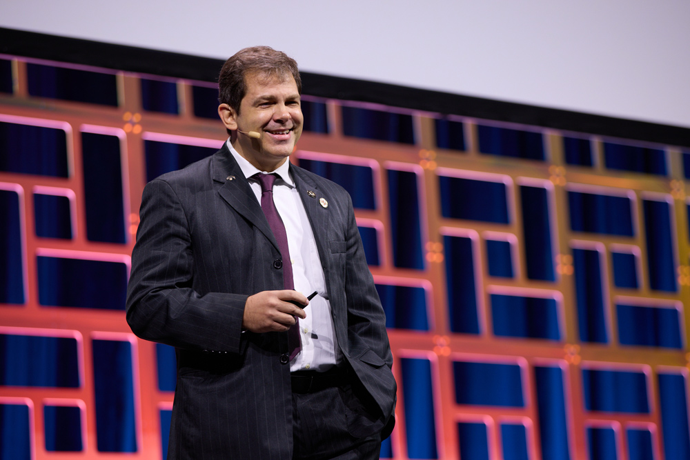
Renato Ambrósio Jr., MD, PhD, presented the Richard L. Lindstrom, MD, Lecture at the 2024 ASCRS Annual Meeting, calling it “one of the highlights of my career.” He began the lecture by sharing quotes from other ophthalmologists about the impact that Dr. Lindstrom has had on the ophthalmic field.
We are celebrating 50 years of ASCRS, he said, noting that the 25th anniversary meeting was the second ASCRS Annual Meeting that he attended, in Seattle, Washington. Before the meeting, he visited the University of Washington, where he met Steven Wilson, MD, who he ultimately trained under. “Dr. Wilson taught me to be a clinician-scientist,” he said. “I see myself today as a refractive surgeon-scientist.”
Source: ASCRS
Dr. Ambrósio’s lecture focused on refractive surgery, specifically highlighting the concept of going “beyond.” Refractive surgery evolved to become a scientific subspecialty of ophthalmology, focusing on elective procedures that aim for refractive correction. But what is the goal? Refractive surgery aims to provide patient satisfaction by improving quality of life, and quality of vision is fundamental along with patient expectations. We have to value and enhance patient education, he said. These excellence standards are based on three major aspects: technology, knowledge, and patient care.
We should consider that refractive surgery should eventually evolve into a specialty medicine with ophthalmology as a prerequisite, Dr. Ambrósio said, mentioning the World College of Refractive Surgery & Visual Sciences. Also, these are not solely elective procedures but involve therapeutic approaches for different diseases, such as keratoconus. “I think about vision and optics optimization every time I see a refractive patient,” he said.
It’s foremost that we customize or individualize the best approach for each eye of each patient, Dr. Ambrósio said, mentioning that along with optimizing the efficacy, each case should be planned to prevent complications like progressive keratectasia, tear dysfunction/ocular pain, interface epithelialization, quality of vision symptoms, and over/under sizing phakic IOLs.
Dr. Ambrósio expanded on the “beyond” theme to discuss imaging in refractive surgery. We started talking about multimodal corneal imaging but demonstrated the concept of multimodal refractive diagnostics. He highlighted several modalities of tests since anamnesis, slit lamp biomicroscopy, corneal topography (Placido), corneal and anterior segment tomography with Scheimpflug and OCT, high-frequency ultrasound, ocular wavefront, axial length, ocular surface imaging, specular and confocal microscopy, and molecular biology and genetics. Considering the plethora of data, he mentioned the role of artificial intelligence in data integration and enhancing clinical decisions.
Physicians should talk to the patient before everything to understand what the patient feels and needs, and this is where patient questionnaires can be valuable.
“We are all here to learn to continuously evolve toward the best version of ourselves.”
Renato Ambrósio Jr., MD, PhD, from a poem he wrote in 2021
He cited work by Stephen Klyce, PhD, on using neural networks for interpreting Placido disc-based topography as an example of a pioneer application of artificial intelligence in medicine.1 He expanded the concept of assessing ectasia risk before refractive corneal procedures, evolving from screening and detecting mild forms of keratoconus and ectatic corneal disease to characterizing the susceptibility for ectasia progression. He said that 1998 was a pioneer year for ectasia because of the first reports of ectasia after LASIK by Theo Seiler, MD, PhD, which alluded to the two-hit pathophysiology of ectasia: the innate properties and the environmental impact. He noted screening, characterization of ectasia susceptibility, confirming the diagnosis, staging, and classifying. Dr. Ambrósio mentioned the Violet June Global Keratoconus Awareness Campaign2 he initiated and said that one of the most important ways to avoid ectasia is to tell patients not to rub their eyes.
He emphasized that it is essential to go beyond, not over, front surface topography. The quest is to characterize the cornea’s susceptibility to developing ectasia with enhanced multimodal diagnostics. Considering that we aim to detect keratoconus and ectasia early, we must include biomechanical assessment and molecular biology.
Dr. Ambrósio cited his American Ophthalmological Society thesis, published in July 2023. The integration of Scheimpflug tomography and biomechanics with the optimized Tomography and Biomechanical Index provides the most accurate approach, considering the 84.4% sensitivity for detecting abnormality among 551 normal topography eyes from patients with very asymmetric ectasia, with 90.1% specificity.3
Also, “beyond” ectasia susceptibility characterization, Dr. Ambrósio highlighted the role of understanding the biomechanical impact with artificial intelligence to quantify the impact of laser vision correction in a relational thickness-altered. The integration of ectasia susceptibility and the impact is further characterized in the enhanced ectasia susceptibility score, which will be available on the BrAIN (Brazilian Artificial Intelligence Networking in Medicine). He mentioned the work by Roger Zaldivar, MD, using artificial intelligence to improve the planning for sizing to predict the postoperative vault of the ICL and the anterior chamber angle.
He referred to the Steinert Lecture at the 2023 ASCRS Annual Meeting given by Vance Thompson, MD. Dr. Thompson asked the question, “What is the difference between standard cataract surgery and refractive cataract surgery?” Dr. Ambrósio responded that the difference is refractive planning. Furthermore, ectasia assessment is relevant for refractive cataract surgery because it impacts the accuracy of IOL power calculation, the quality of vision after premium IOLs, and the safety and efficacy of secondary corneal laser vision correction.
“We are looking at a revolution in the evolution of OCT, and I think it’s crucial to distinguish corneal OCT and anterior segment OCT. There are many machines available and great opportunities for artificial intelligence,” he said. He highlighted the Pentacam Corneal OCT (Oculus), which combines rotating Scheimpflug and ultra high-definition OCT. There is a synergism between Scheimpflug imaging to quantify the amount of opacity and OCT to detail and qualify the layers and the depth of the opacity so that we can plan how to handle those with therapeutic procedures.
He concluded by thanking ASCRS for the great honor of giving the lecture and receiving the award from Dr. Lindstrom. He said that refractive surgery is a mindset that is summarized in a poem that he wrote in 2021: “We are all here to learn to continuously evolve toward the best version of ourselves.”
References
- Maeda N, et al. Neural network classification of corneal topography. Preliminary demonstration. Invest Ophthalmol Vis Sci. 1995;36:1327–1335.
- Ambrósio R. Violet June: The Global Keratoconus Awareness Campaign. Ophthalmol Ther. 2020;9:685–688.
- Ambrósio R, et al. Optimized artificial intelligence for enhanced ectasia detection using Scheimpflug-based corneal tomography and biomechanical data. Am J Ophthalmol. 2023;251:126–142.
Relevant disclosures
Ambrósio: Alcon, Carl Zeiss Meditec, Mediphacos, Oculus, physician CEO of BrAIN
Contact
Ambrósio: dr.renatoambrosio@gmail.com