Refractive
Fall 2024
by Ellen Stodolas
Editorial Co-Director
Epithelial mapping has rapidly gained attention over the last decade with the advent of high-resolution diagnostic devices, specifically high-resolution OCT and ultrasound biomicroscopy (UBM), each of which offer unique advantages.
OCT, said George Waring IV, MD, is more readily available commercially and more efficient in the clinic and provides ease of use for both staff and patients. Ultrasound is less susceptible to optical artifacts, which has additional advantages in terms of reliability of scans and less optical noise. The normative data has been well described at this point, he said, as well as epithelial mapping software that’s been validated for both types of diagnostics.

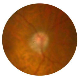
Source: David Huang, MD, PhD, and Yan Li, PhD
“We’ve participated in the early research in epithelial mapping with high-resolution OCT, and this was a big part of our early studies for crosslinking,” Dr. Waring said, adding that it also helped with understanding the natural history of keratometric normalization after epithelial removal in an irregular stromal surface curvature.
Dr. Waring mentioned the early research on understanding the role of epithelial mapping in myopic LASIK and the variation in central thickness epithelial maps that correlate with higher ablation profiles and their potential impact on residual refractive error.1,2 Dr. Waring also mentioned David Huang, MD, PhD, inventor of OCT, and his thesis on being able to discern contact lens warpage and ectasia through epithelial mapping software with a high degree of sensitivity and specificity, and said Dan Reinstein, MD, should be credited for the development of high-resolution ultrasound and application of it for corneal- and lens-based refractive surgery. Dr. Reinstein has done extensive research3–13 on the topic.
With all of these advancements and developments, Dr. Waring said the jury is still out on the universal application of epithelial mapping and its understanding relating to residual refractive error. As it relates to predicting and adjusting for variation in epithelial thickness in refractive enhancements, Dr. Waring said this can apply to post-LASIK patients, or any corneal refractive procedure patient, where the corneal curvature has been changed.
There is a lot of interest in being able to better predict refractive outcomes in the post-corneal refractive state where the epithelium is non-uniform, Dr. Waring said. Multiple attempts have been made to try to predict refractive outcomes in the setting of non-uniform epithelium, and uses for this would be in the post-myopic or post-hyperopic LASIK patient who is interested in an enhancement or the post-myopic LASIK patient who had an IOL now with residual refractive error.
“If we were to unmask the non-uniform epithelium, that may have some variation and decrease predictability in our refractive planning as we’ve decoupled the masking of the non-uniform epithelium in a way we can’t entirely correlate with refraction and therefore the correction of residual refractive error,” Dr. Waring said. “Our opinion is that, although we routinely perform epithelial mapping and study it, we keep this in mind in our planning and in setting expectations with our patients and documenting these expectations about the unpredictability.”
Dr. Waring doesn’t think there is enough data to support the refractive adjustments in planning based on epithelial mapping alone. “We think that there could be a role for further predictive analysis perhaps with artificial intelligence and finite element modeling on the refractive implications,” he said. “However, this is a dynamic circumstance where the epithelium is going to attempt to normalize the corneal curvature irrespective of the prediction.”
Dr. Huang said epithelial mapping can be helpful for the post-cataract surgery patient who might have a good refractive outcome and have residual myopia, hyperopia, or astigmatism. “I think in that situation, before you do any corrective procedure, you want to make sure the cornea has stabilized in terms of edema by following the pachymetric map and in terms of epithelial remodeling by following the epithelial thickness map.” The shape of the cornea can often change slightly because of the corneal incision; the epithelium will adjust to that, and that takes a while to settle down.
Dr. Huang said that an epithelial map can show that dry eye will often have an uneven distribution. There can be more exaggerated thinning superiorly due to the friction from the eyelid, and that can introduce coma, aberration, and apparent astigmatism. If that’s the case, you can increase lubrication and allow time for the corneal epithelium to recover before doing any procedure to correct the astigmatism.
In the case of residual refractive error following cataract surgery, Dr. Reinstein said it’s important to identify whether the visual complaints from the patient are actually the result of residual refractive error or due to the poor contrast and/or dysphotopsia associated with trifocal or multifocal IOLs. “Our plan of action would be to identify the root cause of the visual complaint,” he said. “In addition to the refraction and ocular examination, we use a range of diagnostic tests measuring the epithelium, topography/tomography, and visual quality measurements such as ocular scatter index, contrast sensitivity, light disturbance analyzer, Osiris aberrometer [CSO], subjective and objective point spread function, C-Quant [Oculus], and QoV questionnaire.”
Provided that the patient has good quality of vision following the premium implant and if the cornea appears normal, Dr. Reinstein said LASIK would be the first-choice method to treat the residual refractive error in the majority of cases. “When screening for refractive surgery, every patient will undergo full topography and tomography including epithelial thickness mapping,” he said. “If indicated, the patient will also have an ultrasound measurement of the epithelium using Insight 100 very high frequency digital ultrasound [ArcScan]. In cases where the patient has undergone lens surgery with a premium implant, using a repeatable and accurate refraction technique is key. It is important that the refraction is also repeated on a separate date to ensure consistency of the results.”
Dr. Reinstein stressed that it is important that refractive surgeons understand the importance of preoperative epithelial thickness and the changes that can occur after myopic or hyperopic refractive surgery. “In my opinion, surgeons should not be performing refractive surgery without accurate and repeatable epithelial mapping,” he said. “Most of the newer OCT devices have epithelial mapping function, which has made epithelial mapping more accessible. In our clinic all patients have their epithelium measured as standard at every preop and postop appointment.” He added that epithelial mapping is important following intraocular surgery, as epithelial sloughing during surgery can cause irregularities in the epithelial surface that manifest as increased refractive astigmatism. He also noted the other applications of epithelial mapping, which include keratoconus screening, dry eye assessment, EBMD evaluation, identification of contact lens warpage, and post-refractive surgery where the epithelial remodeling will alter the refractive measurements, which is especially important in hyperopic eyes.
LASIK may have advantages over PRK in these patients, he said, as LASIK has been demonstrated to be more accurate, and the shorter recovery time results in a faster visual recovery. PRK should be reserved for cases where medically indicated, he added.
Dr. Waring also addressed the question of if it would be more favorable to do a LASIK enhancement or a PRK enhancement. One thought is that if we perform LASIK, we are now accounting for the non-uniform epithelium and altering it potentially much less, he said, adding that this makes sense, and there is literature to support this.14 “However, we also think that more often patients are better suited for PRK enhancement in a post-refractive state,” Dr. Waring said. If they’ve already had LASIK, that’s typically where you run into issues. Most of the time, patients do well with a PRK enhancement in these circumstances, he said, and there are additional potential benefits in form fruste or subclinical epithelial basement changes, micro striae, or with subtle or superficial flap irregularities. It can often be therapeutic in a surface ablation treatment, as well as to normalize the corneal surface and shape. “We think there can be an appropriate role for surface ablation in these circumstances. With that said, we also see an opportunity to refine outcomes and prediction factors if we can crack the code on predictive modeling and adjustments for variable epithelial thickness in the post-refractive state.”
Dr. Huang said that he finds both LASIK and PRK effective when there is residual refractive error, and it depends on surgeon preference. Sometimes you may have a scar or previous refractive surgery that makes the cornea irregular, and transepithelial PTK/PRK can improve topographic regularity. “The epithelium can serve as an excellent smoothing agent. I often use transepithelial ablation to improve the postop topography and reduce the postop aberrations, and that helps improve the best corrected visual acuity,” he said. “However, the surgeon should look at the epithelial map to see if the epithelium is masking the irregularity or creating the irregularity. If you have primary epithelial deformation due to dry eye or eyelid effect, transepithelial ablation should be avoided—it would exacerbate the irregularity.”
Discussing epithelial mapping technology, Dr. Huang primarily uses the Visionix (formerly Optovue) Avanti or Solix technology. Both have epithelial mapping capability. “I mainly use it to distinguish between secondary epithelial modulation and primary epithelial deformation. In secondary epithelial modulation, the epithelium becomes thinner to compensate for areas where the anterior stromal surface is steeper or more elevated due to ectasia, scar, deposits, or dystrophies. In primary epithelial deformation, the underlying stroma is smooth, but the epithelial layer is irregular due to lid wiper effect, dry eye, or contact lens warpage. It is important to distinguish between these two disease processes,” Dr. Huang said.
Dr. Huang, and his colleague, Yan Li, PhD, also mentioned several technologies that offer epithelial mapping: MS-39 (CSO), ANTERION (Heidelberg Engineering), and CIRRUS HD-OCT (Carl Zeiss Meditec).
Dr. Reinstein noted a recently published paper15 that found continued improvement and wider implementation of epithelial thickness mapping in OCT devices. He thinks that refractive surgeons should be using epithelial thickness mapping as a tool for all preoperative screening and postoperative assessment and added that it can be easily incorporated as part of the process. “Following refractive surgery, the same preoperative scans can be repeated postoperatively; this is especially useful, as difference maps can then be plotted to monitor progress,” he said.
For those just getting started in epithelial mapping, Dr. Reinstein said it’s important that epithelial maps are used within a multimodal approach in conjunction with other imaging devices in order to build a full picture of the cornea, especially in keratoconus screening. “I would advise liaising with the device manufacturer to ensure that the most optimal scales and color settings are used to be able to detect small irregularities in the cornea,” he said. “Difference maps should also be regularly used and are especially useful in detecting subclinical changes in the epithelium.”
About the physicians
David Huang, MD, PhD
Wold Family Endowed Chair in Ophthalmic Imaging
Professor of Ophthalmology and Biomedical Engineering
Casey Eye Institute
Oregon Health & Science University
Portland, Oregon
Dan Reinstein, MD, MA(Cantab)
London Vision Clinic
EuroEyes Group
London, U.K.
George Waring IV, MD
Waring Vision Institute
Mt. Pleasant, South Carolina
References
- Prospero Ponce CM, et al. Central and peripheral corneal thickness measured with optical coherence tomography, Scheimpflug imaging, and ultrasound pachymetry in normal, keratoconus-suspect, and post-laser in situ keratomileusis eyes. J Cataract Refract Surg. 2009;35:1055–1062.
- Rocha KM, Krueger RR. Spectral-domain optical coherence tomography epithelial and flap thickness mapping in femtosecond laser-assisted in situ keratomileusis. Am J Ophthalmol. 2014;158:293–301.
- Reinstein DZ, et al. High-frequency ultrasound measurement of the thickness of the corneal epithelium. Refract Corneal Surg. 1993;9:385–387.
- Reinstein DZ, et al. Arc-scanning very high-frequency digital ultrasound for 3D pachymetric mapping of the corneal epithelium and stroma in laser in situ keratomileusis. J Refract Surg. 2000;16:414–430.
- Reinstein DZ, et al. Epithelial thickness in the normal cornea: three-dimensional display with Artemis very high-frequency digital ultrasound. J Refract Surg. 2008;24:571–581.
- Reinstein DZ, et al. Corneal pachymetric topography. Ophthalmology. 1994;101:432–438.
- Reinstein DZ, et al. Corneal epithelial thickness profile in the diagnosis of keratoconus. J Refract Surg. 2009;25:604–610.
- Reinstein DZ, et al. Epithelial and corneal thickness measurements by high-frequency ultrasound digital signal processing. Ophthalmology. 1994;101:140–146.
- Reinstein DZ, et al. Comparison of corneal epithelial thickness measurement between Fourier-domain OCT and very high-frequency digital ultrasound. J Refract Surg. 2015;31:438–445.
- Reinstein DZ, et al. Epithelial, stromal, and total corneal thickness in keratoconus: three-dimensional display with Artemis very-high frequency digital ultrasound. J Refract Surg. 2010;26:259–271.
References (continued) - Reinstein DZ, et al. Change in epithelial thickness profile 24 hours and longitudinally for 1 year after myopic LASIK: three-dimensional display with Artemis very high-frequency digital ultrasound. J Refract Surg. 2012;28:195–201.
- Reinstein DZ, et al. LASIK for the correction of high hyperopic astigmatism with epithelial thickness monitoring. J Refract Surg. 2017;33:314–321.
- Reinstein DZ, et al. Transepithelial phototherapeutic keratectomy protocol for treating irregular astigmatism based on population epithelial thickness measurements by Artemis very high-frequency digital ultrasound. J Refract Surg. 2014;30:380–387.
- Rohlf D, et al. Outcomes of LASIK vs PRK enhancement in eyes with prior cataract surgery. J Cataract Refract Surg. 2023;49:62–68.
- Reinstein DZ, et al. Epithelial thickness mapping for corneal refractive surgery. Curr Opin Ophthalmol. 2022;33:258–268.
Relevant disclosures
Huang: Canon, Cylite, Genentech, Intalight, Visionix
Reinstein: ArcScan, Carl Zeiss Meditec, CSO
Waring: None
Contact
Huang: huangd@ohsu.edu
Reinstein: dzr@londonvisionclinic.com
Waring: georgewaringiv@gmail.com