ASCRS News: EyeWorld Journal Club
September 2022
by Anthony Mai, MD, Mike Murri, MD, and Jeff Pettey, MD, MBA

Residency
Program Director
Moran Eye Center
University of Utah
Salt Lake City, Utah
Laser-assisted in situ keratomileusis (LASIK) has become the refractive procedure of choice over the past few decades. LASIK enhancements are commonly performed for residual refractive errors, regression, or surgically induced astigmatism.1 Enhancement is done by direct surface ablation, cutting a new flap, or relifting the old flap. Direct surface ablation has increased risk for pain, infection, longer recovery time, and corneal haze.2,3,4,5 Recutting may cause a free cap, button hole, ordisplaced slivers of stromal tissue.7,8,9 Compared to recutting, relifting may yield fewer complications and better long-term stability of refractive error and visual acuity.7,10 Although flap relifting years after LASIK has been questioned, recent studies show success more than 15 years after the initial procedure.6,11 Nevertheless, relifting can be technically challenging and complicated by epithelial ingrowth (EI).12,13 Prior studies have suggested pre-enhancement time interval, microkeratome use, loose epithelium, and advanced age to be possible post-relifting EI risk factors.14,15,16,17,18 With this current study, Chang et al. seek not only to confirm the viability of flap relifting years after LASIK but also to assess therelationship between relifting success and EI with pre-enhancement time interval, age during relift, sex, and primary LASIK flap creation method.

Resident
Moran Eye Center
University of Utah
Salt Lake City, Utah
Methods
The authors retrospectively reviewed all LASIK relifting enhancement cases performed by one surgeon at the Hong Kong Sanatorium & Hospital between 1997 and 2019. They included data only on the first relift procedure if a patient had multiple. They excluded patients with second flap-related procedures within 75 days of the first relift, less than 75 days of follow-up in the absence of epithelial ingrowth, macular disease, insufficient stromal bed, evidence or suspicion of keratectasia, hypertension, history or risk of retinal vessel occlusion, glaucoma, and other procedures like corneal crosslinking. Their primary outcomes were relifting success, EI development, intraoperative complications, and postoperative corrected distance visual acuity (CDVA). They defined clinically significant EI as distance visual acuity (corrected or uncorrected) loss, foreign body sensation, keratolysis, epithelial irregularity, or flap revision desired by either the surgeon or the patient. The study performed statistical analysis with R, especially using binary logistic regression to evaluate the association between relifting success and EI with pre-enhancement time interval, age during relift, sex, and primary LASIK flap creation method. These associations were presented as odds ratios.
The authors described the relifting procedure used in the study. In summary, the previous flap edge was visualized under a slit lamp, anesthetized by 0.5% proparacaine, and initially lifted by a 25-gauge needle bent 2 mm from the tip that extended 1.5 mm inward. The needle created a 2–3-mm long flap lift that was
pressed back on the stromal bed. The Seibel II IntraLase flap lifter and retreatment spatula short end extended the incision along the old flap’s circumference, after which the long end lifted the flap. The stromal bed was then treated by an excimer laser and scrubbed by a Merocel sponge. A feeding tube connected to suction removed residual fluid under the flap, and a bandage contact lens was placed.
Resident
Moran Eye Center
University of Utah
Salt Lake City, Utah
Results
The authors included 73 eyes from 68 patients. The mean follow-up periods were 4.3±4.7 years. The mean time interval between LASIK and relift was 8.6±6.4 years. Relifting was successfully performed in 46 eyes (63%) >5 years and in 34 eyes (47%) >10 years after primary LASIK. Relifting was successful in 71 eyes (97.3%). The 2 eyes with failed relifting had successful recutting procedures without complications. Of the successfully relifted flaps, 12 (16.9%) developed EI. Of these, 3 (4.2%) were clinically significant. None of these lost CDVA after EI removal. EI only recurred in 1 of those 3 eyes but was clinically insignificant. Seven eyes (9%) lost one line of CDVA but no eyes lost >1 line. The association between pre-enhancement time intervals with relift success or EI development was not statistically significant, even after adjusting for patient characteristics like primary LASIK flap creation method, year of relift, or age at relift. There were also no direct associations found between relift success or EI development with sex, patient age at relift, year of relift, and primary LASIK flap creation method (microkeratome vs. femtosecond laser).
Discussion
Various studies have advised against relifting a LASIK flap after the 1-year postop mark.12,19 However, conflicting reports have demonstrated successful flap relifting after this time period, even up to 15–20 years after surgery.6,11 Before considering LASIK enhancement complications, the flap must first be successfully mobilized and lifted. This study reported a success rate of 97% with half of its cases occurring more than 10 years after primary LASIK surgery at a maximum of 22 years later.
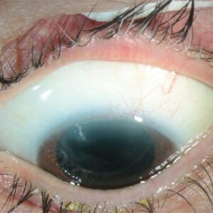
The feared complication of EI occurred in roughly 17% of patients, which is consistent with previously reported numbers of 0 to 18%.17,20 However, clinically significant EI occurred in only 4.2% of relifted flaps, none of which lost CDVA after treatment. Notably, a greater pre-enhancement time interval was not associated with increased EI. This study also could not confirm the previously suggested association between patient age and EI.21 This study suggests that loose epithelium may have been associated with epithelial ingrowth. However, this is often not clearly documented and needs further study.
A microkeratome flap may theoretically have a higher EI risk compared to a femtosecond laser flap due to the >90-degree side cut angle. This same attribute may also decrease scarring and increase the ease of flap relift compared to a femtosecond laser flap. Interestingly, there were no significant differences in relift success and EI incidence between the two tools. The flap relift method itself may also influence EI incidence. The authors were concerned that using a probe at 180 degrees from the hinge and lifting the rest of the flap with forceps may lead to greater rates of EI. Therefore, they instead created an opening at the flap margin with a sharp needle into which a spatula was used to cleave and lift the flap. They hoped this method would decrease the number of epithelial cells introduced under the flap.
The study is limited by short follow-up, poor documentation of loose epithelium prior to relifting, and lack of a clear definition of EI with which to compare with other studies. It is also limited by sample size factors that made comparison of variables (sex, flap creation method, successful vs. unsuccessful lifts) difficult. Lastly, this study’s inclusion of all EI cases makes it appear to have a higher EI incidence compared to other papers that only report clinically significant EI.
Conclusion
According to this study, LASIK enhancement via flap relifting can be safe years after primary LASIK treatment. It may have fewer complications compared to alternatives like surface ablation or recutting. Clinically significant EI is rare and does not cause vision changes after it was removed. The authors also showed that previously suggested risk factors for EI like pre-enhancement time interval, flap creation tool, sex, and age were unsupported. This is the largest retrospective study to date that examines LASIK flap relifting and describes a specific surgical technique with low complication rates. The authors present a compelling argument for flap relifting to become the preferred method for LASIK enhancement many years after primary treatment.
Effect of time since primary laser-assisted in situ keratomileusis on flap relift success and epithelial ingrowth risk
Chang JSM, et al.
J Cataract Refract Surg. 2022;48:705–709.
- Purpose: To assess the association of time since primary laser-assisted in situ keratomileusis (LASIK) with flap relift success and risk for epithelial ingrowth (EI) in eyes undergoing flap relift after primary LASIK
- Setting: Hong Kong Sanatorium & Hospital, Hong Kong Special Administrative Region
- Design: Retrospective observational case series
- Methods: 73 eyes with flaps relifted for refractive enhancement LASIK were included. Main outcome measures included rate of relift success and EI; associations of time since primary LASIK, sex, age at relift, year of relift, and flap creation method in primary LASIK with relift success and EI
- Results: Of the 73 eyes included, relifting was successful in 71 eyes (97.3%). Among the successfully relifted eyes, 12 (16.9%) developed EI, of which 3 (4.2%) were clinically significant. No eyes lost more than 1 line of corrected distance visual acuity. The time since primary LASIK (up to 22 years), sex, age at relift, year of relift, and flap creation method in primary LASIK were not associated with relift success or EI.
- Conclusion: With the described surgical technique, flaps could be successfully relifted without much difficulty up to 22 years after primary LASIK with a low incidence of EI.
References
- Moshirfar M, et al. LASIK enhancement: clinical and surgical management. J Refract Surg. 2017;33:116–127.
- Cagil N, et al. Effectiveness of laser-assisted subepithelial keratectomy to treat residual refractive errors after laser in situ keratomileusis. J Cataract Refract Surg. 2007;33:642–647.
- Carones F, et al. Evaluation of photorefractive keratectomy retreatments after regressed myopic laser in situ keratomileusis. Ophthalmology. 2001;108:1732–1737.
- de Rojas V, et al. Infectious keratitis in 18,651 laser surface ablation procedures. J Cataract Refract Surg. 2011;37:1822–1831.
- Schallhorn SC, et al. Flap lift and photorefractive keratectomy enhancements after primary laser in situ keratomileusis using a wavefront-guided ablation profile: Refractive and visual outcomes. J Cataract Refract Surg. 2015;41:2501–2512.
- Caster AI. Flap-lift LASIK 10 or more years after primary LASIK. J Refract Surg. 2018;34:604–609.
- Davis EA, et al. Lasik enhancements: a comparison of lifting to recutting the flap. Ophthalmology. 2002;109:2308–2314.
- Rubinfeld RS, et al. To lift or recut: changing trends in LASIK enhancement. J Cataract Refract Surg. 2003;29:2306–2317.
- Vaddavalli PK, et al. Complications of femtosecond laser-assisted re-treatment for residual refractive errors after LASIK. J Refract Surg. 2013;29:577–580.
- Domniz Y, et al. Recutting the cornea versus lifting the flap: comparison of two enhancement techniques following laser in situ keratomileusis. J Refract Surg. 2001;17:505–510.
- Alió Del Barrio JL, et al. Laser flap enhancement 5 to 9 years and 10 or more years after laser in situ keratomileusis: Safety and efficacy. J Cataract Refract Surg. 2019;45:1463–1469.
- Santhiago MR, et al. Flap relift for retreatment after femtosecond laser-assisted LASIK. J Refract Surg. 2012;28:482–487.
- Randleman JB, Shah RD. LASIK interface complications: etiology, management, and outcomes. J Refract Surg. 2012;28:575–586.
- Wang MY, Maloney RK. Epithelial ingrowth after laser in situ keratomileusis. Am J Ophthalmol. 2000;129:746–751.
- Letko E, et al. Influence of original flap creation method on incidence of epithelial ingrowth after LASIK retreatment. J Refract Surg. 2009;25:1039–1041.
- Caster AI, et al. Incidence of epithelial ingrowth in primary and retreatment laser in situ keratomileusis. J Cataract Refract Surg. 2010;36:97–101.
- Henry CR, et al. Epithelial ingrowth after LASIK: clinical characteristics, risk factors, and visual outcomes in patients requiring flap lift. J Refract Surg. 2012;28:488–492.
- Ting DSJ, et al. Epithelial ingrowth following laser in situ keratomileusis (LASIK): prevalence, risk factors, management and visual outcomes. BMJ Open Ophthalmol. 2018;3:e000133.
- Durrie DS, Aziz AA. Lift-flap retreatment after laser in situ keratomileusis. J Refract Surg. 1999;15:150–153.
- Walker MB, Wilson SE. Incidence and prevention of epithelial growth within the interface after laser in situ keratomileusis. Cornea. 2000;19:170–173.
- Chan CCK, Boxer Wachler BS. Comparison of the effects of LASIK retreatment techniques on epithelial ingrowth rates. Ophthalmology. 2007;114:640–642.
Contact
Mai: Anthony.Mai@nick-mamalishsc-utah-edu
Murri: Mike.Murri@nick-mamalishsc-utah-edu
Pettey: jeff.pettey@hsc.utah.edu