ONLINE EXCLUSIVE
June 2022
by Liz Hillman
Editorial Co-Director
“It’s alive!” —Victor Frankenstein, in James Whale’s 1931 film “Frankenstein”
Investigators from the John A. Moran Eye Center at the University of Utah, with collaboration from Scripps Research, have revived photoreceptor cells from organ donor eyes and restored communication between them. The research, according to a press release from the Moran Eye Center, “[stands] to transform brain and vision research.”
Fatima Abbas, PhD, lead author of the research, which was published in May in the journal Nature,1 said in the press release that the team was able to “wake up photoreceptor cells in the human macula” from eyes that were obtained up to 5 hours after the organ donor’s death. Dr. Abbas said the cells “responded to bright light, colored lights, and even very dim flashes of light.”
The authors wrote in the Nature paper that while some human organs can be successfully transplanted from deceased donors, “tissues from the central nervous system rapidly lose viability after circulation ceasesimpeding their potential for transplantation.” The paper further described how the investigators “systemically [examined] the kinetics of death and neuronal revival.”
Source: John A. Moran Eye Center at the University of Utah
In the research, the investigators found that while photoreceptor cells could be revived up to 5 hours after the donor’s death, the cells no longer communicated with each other. They identified oxygen deprivation as a major reason leading to the loss of this ability. From here, the team obtained donor eyes less than 20 minutes after death and transported them in a unit specially designed to restore oxygen and other nutrients, the Moran press release described.
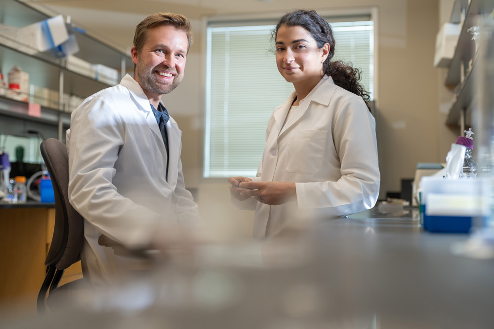
Frans Vinberg, PhD, designed the donor transport unit and built a system that stimulated the retina and measured electrical activity in the cells. Electrical signals, B waves, like those measurable in living eyes were restored in the “retinal patches,” as described in a video interview about the research.
“We were able to make the retinal cells talk to each other, the way they do in the living eye to mediate human vision,” Dr. Vinberg said in the press release. “Past studies have restored very limited electrical activity in organ donor eyes, but this has never been achieved in the macula, and never to the extent we have now demonstrated.”
Source: John A. Moran Eye Center
According to Moran, this process could be used to study other neuronal tissues and neurodegenerative diseases. For vision and therapeutics research, Dr. Vinberg said that these findings could reduce reliance on non-human primates and animal models that don’t as accurately model the human eye (such as mice, which don’t have a macula).
“The scientific community can now study human vision in ways that just aren’t possible with laboratory animals,” Dr. Vinberg said.
Anne Hanneken, MD, a retina specialist involved with the research, said in the press release that “until now, it hasn’t been possible to get the cells in all of the different layers of the central retina to communicate with each other the way they normally do in a living retina.
“Going forward, we’ll be able to use this approach to develop treatments to improve vision and light signaling in eyes with macular diseases, such as age-related macular degeneration,” she said.
For more, read the full press release and the paper published in Nature.
Reference
- Abbas F, et al. Revival of light signalling in the postmortem mouse and human retina. Nature. 2022. Online ahead of print.
About the researchers
Fatima Abbas, PhD
Postdoctoral Researcher
John A. Moran Eye Center
University of Utah
Salt Lake City, Utah
Anne Hanneken, MD
Associate Professor of Molecular Medicine
Department of Molecular Medicine
Scripps Research
Retina Consultants of San Diego
La Jolla, California
Frans Vinberg, PhD
Assistant Professor
Department of Ophthalmology and Visual Sciences
John A. Moran Eye Center
University of Utah
Salt Lake City, Utah