Glaucoma
September 2022
by Ellen Stodola
Editorial Co-Director
IOP elevation following intravitreal injections can be divided into short-term and long-term elevations, said Christina Y. Weng, MD, MBA. Short-term elevations are common, and some studies have estimated that they affect greater than 90% of patients. “Research, as well as personal experience, has shown that IOP can often elevate to the 30s or even 40s mm Hg for a transient period of time immediately following injection of anti-VEGF agents,” Dr. Weng said. “However, these elevations are short lasting and often resolve within 30–60 minutes, although there are some reports that IOP can remain elevated for more than a day.” She added that intravitreal injections of steroid agents may be the caveat to this because in addition to the initial IOP elevation, they can sometimes lead to elevations that occur 4–6 weeks after the injection.

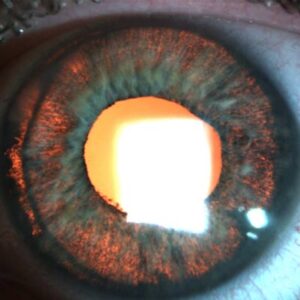
Source: Christina Y. Weng, MD, MBA
Dr. Weng said that the data becomes “a bit more ambiguous” with long-term elevations, and there have been a number of studies with varying conclusions. “However, if you look at the largest meta-analyses and database studies, the takeaway is that persistent IOP elevation or glaucoma after repeated intravitreal injections can occur but is rare,” she said.
Malik Y. Kahook, MD, noted the incidence of IOP elevations is about 1–3% of patients post-anti-VEGF use. “The number is not that high, and it is still unclear to me if we see it more with one anti-VEGF agent over another,” he said. “I think some of the information on IOP elevations post-injection of this class of medication has been conflated by observers,” he said.
He added that there were many reports years ago, including from his group at the University of Colorado, that were focused on case clusters of IOP elevation related to compounding pharmacy practices. “We found silicone oil bubbles in repackaged bevacizumab syringes that were related to practices by specific pharmacies and related to shipping-induced mechanical stress on the syringes,” he said.1,2 “I think these events are very different from what is seen in regular practice and with use of FDA-approved versions of anti-VEGF agents. Furthermore, I think compounding pharmacy practices have evolved and are much better now compared to a few years ago.” This has resulted in a lower incidence of IOP elevation post-anti-VEGF agents, in Dr. Kahook’s experience.
Naresh Mandava, MD, has looked at this issue with Dr. Kahook in several studies. Dr. Mandava first became interested in this topic after hearing a talk from a fellow physician who presented a 20% rate of intractable elevation in IOP when injecting bevacizumab. With further exploration into this, Dr. Mandava said he hasn’t seen levels that high, but he shared similar comments as Dr. Kahook about how certain compounding pharmacies seem to have issues, likely due to silicone oil microdroplets in the syringes.
This led to further research, as well as conversations with many large pharmacies. As such, changes have been made to ensure the drug is transferred appropriately. Dr. Mandava added that the way that these syringes were transported on dry ice was also a concern because the drug could freeze and thaw during travel time. “Our multidisciplinary group of clinicians and scientists in ophthalmology and the pharmaceutical sciences was the first to elucidate the cause of intractable elevation of intraocular pressure in patients receiving these therapies,” he said. “This research led to changes in compounding pharmacy practices, which has decreased the incidence of these complications.”
Risk factors
Dr. Weng said that if the patient has ocular hypertension, a diagnosis of glaucoma at baseline, or other risk factors for developing glaucoma, the risk for post-injection IOP elevation is higher. “Ocular characteristics such as lens status and axial length have been explored without definitive correlation established,” she said. “As we age, the sclera becomes increasingly rigid, which could also theoretically contribute to IOP elevation.”
Dr. Weng added that another factor that seems to increase risk is the frequency or cumulative number of injections that a patient has received. With regard to the medication itself, this is unclear. In one IRIS Registry study of 23,776 patients, on average, 2.6% sustained a clinically significant IOP rise with anti-VEGF drugs overall compared to 1.5% in the fellow untreated eye, Dr. Weng said. However, this difference was not observed with the aflibercept subgroup compared to the bevacizumab and ranibizumab subgroups.3
Dr. Kahook said his studies have shown that the incidence of IOP spikes was much higher in patients with pre-existing glaucoma. In one study, they found patients with pre-existing glaucoma experienced higher rates of elevated IOP when compared to patients without pre-existing glaucoma (33% vs. 3.1%, respectively; p<0.001).4 Clinicians should treat patients with extra caution when they require anti-VEGF injections in the setting of co-existing glaucoma and retinal pathology. “Added vigilance to check IOP post-injection and routine check of pressure with Goldmann applanation is key,” he said. “When patients are seen in injection clinics, it is imperative that they receive pressure checks as part of the routine workup, something that is not done across the board.”
Minimizing risk
If there is a patient in whom avoidance of an IOP spike is critical, Dr. Weng said the retina specialist could consider performing a pre-injection anterior chamber paracentesis and prescribing prophylactic IOP-lowering drops. “Additionally, pre- and post-injection IOP measurements as well as close tracking temporally may provide insights into whether a change in treatment frequency or a switch in anti-VEGF agent should be considered.”
Dr. Mandava said he will always check the pressure before doing an injection. If the pressure is mildly elevated, he will move forward. But if it’s elevated into the mid-20s or higher, he thinks about what the cause may be, examines the eye and drainage system, makes sure there are no other issues, and looks at optic nerves to see if there’s any evidence of asymmetry. If it’s above a certain pressure, he may withhold the injection, start the patient on topical medication, and see them back for the injection.
Injections in the setting of MIGS/other glaucoma procedures
For those with a history of MIGS/glaucoma surgery, there is no consensus on whether to modify the injection or peri-injection approach, Dr. Weng said. “I am always mindful of where trabeculectomy blebs or tube shunt plates are located and avoid injecting through these areas, but I do not currently do anything differently for these patients aside from reminding them to continue with regular follow-up with their glaucoma specialist and monitoring their pre-injection IOP each time they come to see me,” she said.
Dr. Weng said she also likes to involve a glaucoma specialist any time she is concerned that an injection-associated IOP elevation is becoming persistent, as they will be best able to assess whether a patient requires surgical intervention. “Fortunately, of the millions of intravitreal injections given each year, only a very small proportion of patients will require a glaucoma procedure as a direct result of injections.”
When IOP spikes occur
Dr. Kahook said that IOP spikes post-anti-VEGF injections come in two forms. “The first is an acute rise that happens in the majority of patients and is related to the expansion of volume within the eye and necessitates time for the eye to reequilibrate,” he said. This can be seen in both healthy and glaucomatous eyes, but it takes longer for eyes with glaucoma to reequilibrate given the compromised aqueous outflow channels.
“The second form of IOP elevation is chronic and can be seen after one or more injections and can last for weeks to months or longer if not treated with medications or surgical intervention,” Dr. Kahook said, adding that it is this second form that results in higher rates of morbidity if not diagnosed and addressed. “This is also the reason that I advocate for making IOP checks part of the routine injection clinic workup before each injection takes place.”
Dr. Weng said spikes are not uncommon immediately following the injection but usually will return to baseline within 30–60 minutes. What is less understood are the late-onset IOP spikes that can occur months or even years after a cumulative number of injections. “With intravitreal steroid injections, you can also see a subacute IOP spike in about one-third of patients that typically occurs 4–6 weeks following the injection, which is why I bring these patients back in that timeframe to check their IOP,” she said.
What to do after an IOP spike
A post-injection IOP elevation will typically resolve on its own within an hour, Dr. Weng reiterated, but if it persists with an IOP >35 mm Hg after a reasonable wait time in the office, she’ll start an IOP-lowering drop and see that patient back again in a few days. If it persists with an IOP >35 mm Hg and the patient has pain or corneal edema due to the elevated IOP, she will consider performing an anterior chamber paracentesis and discharge the patient on IOP-lowering drops. “If the patient has a higher than average risk for glaucoma, I would also consider referring to a glaucoma specialist for baseline evaluation,” she said.
Dr. Weng added that the risk of vision loss from the many diseases that intravitreal injections treat is significantly greater than the small risk of injection-induced glaucoma. “However, there needs to be more research conducted in this area so that we can continue to optimize the safety profile of intravitreal injections, the most common ophthalmic procedure we do today,” she said.
About the physicians
Malik Y. Kahook, MD
Slater Family Endowed Chair in Ophthalmology
University of Colorado Anschutz Medical Campus
Aurora, Colorado
Naresh Mandava, MD
Professor and Chair
Department of Ophthalmology
University of Colorado Anschutz Medical Campus
Aurora, Colorado
Christina Y. Weng, MD, MBA
Professor of Ophthalmology
Cullen Eye Institute
Baylor College of Medicine
Houston, Texas
References
- Liu L, et al. Silicone oil microdroplets and protein aggregates in repackaged bevacizumab and ranibizumab: effects of long-term storage and product mishandling. Invest Ophthalmol Vis Sci. 2011;52:1023–1034.
- Kahook MY, et al. High-molecular-weight aggregates in repackaged bevacizumab. Retina. 2010;30:887–892.
- Atchison EA, et al. The real-world effect of intravitreous anti-vascular endothelial growth factor drugs on intraocular pressure: an analysis using the IRIS Registry. Ophthalmology. 2018;125:676–682.
- Good TJ, et al. Sustained elevation of intraocular pressure after intravitreal injections of anti-VEGF agents. Br J Ophthalmol. 2011;95:1111–1114.
Relevant disclosures
Kahook: Alcon, Aurea Medical, Equinox, Fluent Ophthalmics, Johnson & Johnson Vision, New World Medical, SpyGlass Ophthalmics, SpyGlass Pharma
Mandava: None
Weng: Allergan, Alcon, Alimera Sciences, DORC, Genentech, Novartis, Regeneron, REGENXBIO
Contact
Kahook: malik.kahook@gmail.com
Mandava: Naresh.Mandava@cuanschutz.edu
Weng: Christina.Weng@bcm.edu