Cataract
July 2021
by Liz Hillman
Editorial Co-Director
There are many potential complications cataract surgeons need to prepare for in the OR. One of them is zonulopathy.
“Recognizing zonulopathy and adopting strategies to mitigate zonular damage is critically important not only for short-term success but perhaps more importantly long-term success to reduce the risk of late IOL dislocation,” said D. Brian Kim, MD.


Source (all): D. Brian Kim, MD
Preoperative recognition
Past ocular history such as trauma, retinitis pigmentosa, angle closure glaucoma attack, a family history of ectopia lentis, or history of pseudoexfoliation “should raise your index of suspicion for zonulopathy,” Dr. Kim said. He also said a small pupil coupled with a dense cataract should be treated like a case with loose zonules “until proven otherwise.”
Nicole Fram, MD, said that obvious signs include areas of missing zonules, such as colobomatous changes, Marfan syndrome, pseudoexfoliation with phacodonesis, and traumatic cataract. More subtle signs include pseudoexfoliation in the setting of a shallow chamber, asymmetric shallowing of the AC where the depth changes as the anterior chamber is examined.
“Surgeons should always proceed with caution in patients with pseudoexfoliation, high myopia, history of PPV or trabeculectomy, uveitis or retinopathy of prematurity, aniridia. All of these conditions cause a disturbance of the blood aqueous barrier and can lead to weak zonules,” Dr. Fram said.

Source (all): D. Brian Kim, MD
Intraoperative signs and management
One has to constantly be on the lookout for zonulopathy at various stages in the case, Dr. Kim said. “Unfortunately, patients often may not show signs of zonulopathy until you are in the operating room,” he said.
There might be signs of zonulopathy before even starting your incision. Dr. Fram said if you see more of the edge of the crystalline lens than normal or if there is a jiggle indicative of phacodonesis, these are warning signs of loose zonules.
Dr. Kim said cases with zonulopathy or zonular laxity will see the lens move backward more than usual when filling the anterior chamber with OVD.
Another opportunity to identify zonulopathy is when starting the capsulorhexis.
“There is often striae resembling elephant’s skin, indicating weak zonules,” Dr. Fram said. “In the moment, you can stain with trypan to make sure the rhexis edge can always be identified.”
Dr. Kim described this as the spider sign, which occurs when just puncturing the anterior capsule with forceps for the capsulorhexis.
“The spider sign occurs because the anterior capsule sags backward as the downward force is applied,” he explained.
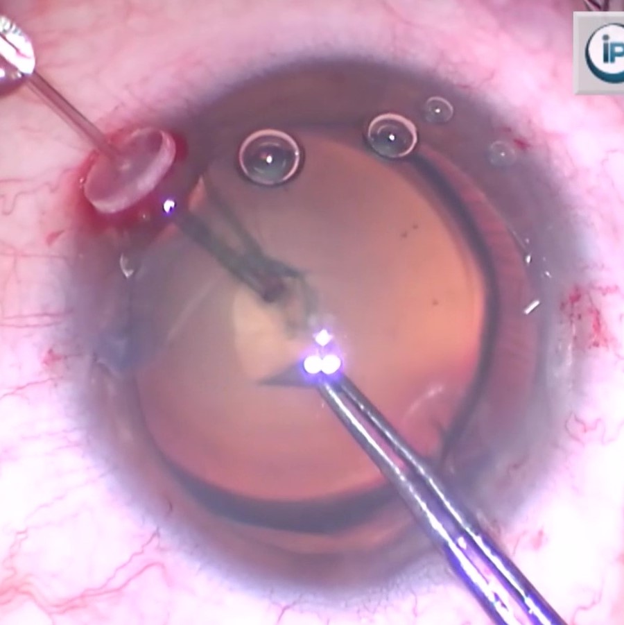
As you’re pulling the rhexis flap, the anterior capsule and bag will move, creating distinctive folds in front of the flap, if the zonules are weak, Dr. Kim continued. He added that capsule retractors can be placed behind the tear to provide counterforce if the rhexis flap doesn’t tear effectively.
With hydrodissection, if the lens doesn’t spin easily with the cannula, this could suggest zonulopathy. Dr. Kim said the surgeon should stop trying to spin the lens, as it could damage zonules further.
When performing phaco, especially with current generation phaco units that can achieve high vacuum, Dr. Kim said any trampolining of the lens would be a sign of zonulopathy.
Other intraoperative signs of zonulopathy, according to Dr. Kim, include the straight line sign, which occurs when there’s striae parallel to the limbus around the peripheral capsular bag. Aggressive I/A in this case can cause the capsular bag to collapse into the I/A tip. In cases of zonulopathy, avoid pulling toward the center with I/A, opting for tangential movement with lower vacuum levels.

Source: D. Brian Kim, MD
Zonulopathy management
Mild zonulopathy can be fairly uneventful in routine cataract surgery, provided key strategies are used to prevent further zonular damage, Dr. Kim said. More severe cases at risk for lens decentration, dislocation, or posterior capsule rupture and dropped lens require more advanced surgical tools and techniques.
“With proper planning one can achieve a successful cataract removal and a well-centered IOL,” Dr. Kim said
Trypan blue can be used to help visualize the capsulorhexis edge, which is important when capsule support devices are needed, Dr. Kim said.
Dr. Kim advised capsule retractors, rather than iris hooks, due to their ability to support the bag, providing a wider base and longer reach for deeper capsular fornix support. He said he would use a capsular tension ring in cases of mild zonulopathy or those with 3 clock hours or less of zonular dehiscence.
“My preferred technique is to use an injector for the CTR with a Sinskey hook. As the CTR is being delivered, the Sinskey hook is used to hook the leading eyelet, and the Sinskey hook holds the leading part of the CTR and reduces torsional stress on the capsular bag and zonules,” Dr. Kim explained.
If zonulopathy is more severe or zonular dehiscence is greater, he said he might use a CTR with extra support like a capsular tension segment with Gore-Tex sutured to the sclera. He said practicing skills in preparation for more advanced cases like this is imperative.
“There are many complex cataract surgery wet labs including at the ASCRS Annual Meeting and the AAO Annual Meeting. There are also model eyes such as the SimulEye [InsEYEt], which is easily accessible and can be practiced in the comfort of your own operating room,” Dr. Kim said.
Once any support devices are placed, Dr. Kim described “zonule-friendly lens disassembly.” This includes using a technique that doesn’t depend on a mobile lens or sculpting.
“I developed double chop and cross chop, which enable the surgeon to divide the lens into smaller pieces without needing to rotate the lens within the capsular bag,”1 Dr. Kim explained. “Learning new skills to disassemble the lens with minimal zonular stress can be a game changer for these situations.”
For IOL selection, Dr. Kim thinks a three-piece IOL is better for these cases.
“If the IOL dislocates in the future, a single- piece acrylic IOL might have to be explanted and replaced, which results in a bigger surgery. In contrast, a three-piece acrylic IOL can be salvaged and secured by lassoing it with Gore-Tex sutured to the sclera,” he said.
Dr. Fram follows a similar technique for milder zonulopathy vs. more severe cases. She said she’ll use capsule retractors, such as Chang Modification (MicroSurgical Technology), and a CTR for less than 3 clock hours of zonulopathy. Ahmed segments (Morcher) and Gore-Tex sutures can be used if the capsulorhexis doesn’t center after CTR placement, she said.
“Another indication for fixation with CTR segments, Cionni ring [Morcher], or modified Malyugin CTR [Morcher] includes diffuse zonulopathy. If the IOL is in the bag and the entire complex is mobile with clear pseudophacodonesis, it is important to fixate the capsule bag to the sclera as the CTR alone may not be a definitive solution. This is particularly important in the case of a planned toric or diffractive/EDOF IOL placement,” Dr. Fram said.
Zonulopathy and FLACS
More modern settings on femtosecond lasers have improved capabilities that Dr. Fram said can have benefits in cataract cases that include zonulopathy. New settings including increased speed of the capsulotomy, flat docking, contact lens interface on the applanation system, liquid interface, and hybrid applanation/liquid interface allow for more stable and stronger capsulotomies, she said.
Dr. Fram thinks the benefits of FLACS for zonulopathy cases are three-fold. First, it creates a perfectly centered and sized capsulotomy, which is important in the setting of shallow chambers and pseudoexfoliation eyes, which are susceptible to capsule contraction. Second, the “pre-chop” fragmentation facilitates easier lens manipulation. Third, pneumodissection from the femtosecond laser bubbles reduce the need for hydrodissection and minimize the need for lens rotation.
“All of these factors improve reproducibility of a complex surgery,” she said, adding that her practice recently evaluated 32 eyes that had FLACS with Chang Modification capsule retractors. In this review, there were two tears in the anterior capsule, both in aniridic eyes, which Dr. Fram said are known to be more friable. Dr. Fram said FLACS capsulotomy should be avoided in aniridic eyes.
Dr. Kim said he doesn’t perform FLACS, but he thinks it has an advantage in creating a perfect capsulotomy and in lens segmentation. One drawback could be that some of the clues of zonulopathy are seen in certain stages of manual cataract surgery and would be lost in FLACS, he noted.
Though he doesn’t have experience with an automated capsulotomy device like Zepto (Centricity Vision) or miLOOP (Carl Zeiss Meditec), Dr. Kim said these tools could be helpful in cases with zonulopathy.
ARTICLE SIDEBAR
What’s new?
Dr. Fram included a few newer concepts and devices in her discussion on zonulopathy. She said there is a concept of a “dead bag,” coined by Samuel Masket, MD. Such capsules have little to no fibrosis and over time will present with zonulopathy and a dislocated IOL-bag complex if the issue occurs near the zonule. Dr. Fram said Liliana Werner, MD, and Nick Mamalis, MD, have been examining specimens of this and have found that the capsules appear to resemble true exfoliation or delamination of the capsule.
“Of note, these capsules do not hold anchoring segments well secondarily, and the best approach is an IOL exchange in these patients,” Dr. Fram said.
She also mentioned two OUS sutured segments that are showing promise: AssiAnchor designed by Ehud Assia, MD, and the Paperclip Capsule Stabilizer designed by Soosan Jacob, MD. Dr. Fram brought up the X1 Iris Speculum (Diamatrix), which, though indicated for iris expansion, has shown in an off-label capacity the ability to stabilize the capsule in cases of zonulopathy.
About the physicians
Nicole Fram, MD
Advanced Vision Care
Los Angeles, California
D. Brian Kim, MD
Private Practice
Professional Eye Associates
Dalton, Georgia
Reference
- Kim DB. Cross chop: Modified rotationless horizontal chop technique for weak zonules. J Cataract Refract Surg. 2009;35:1335–1337.
Relevant disclosures
Fram: None
Kim: None
Contact
Fram: DrFram@avceye.com
Kim: kim@professionaleye.com