For a more recent article on this topic, see “Recognizing, understanding, and treating conjunctivochalasis.”
Opinions and Commentary: eyeConnections
January 2009
by J. E. “Jay” McDonald II, M.D.
Making sense of an unusual occurrence
How many times do I read a post and go, “Wow—I never ever knew.” Well, the case below is a new one for me, and hopefully I was not the only one to hear of this phenomena for the first time. Thanks to all those that post and answer on eyeCONNECT. I continue to expand my knowledge base on a daily basis. Here was the original message that generated the discussion—I’m sharing responses that appeared on both the cataract and refractive listservs.
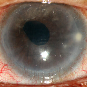
I have a healthy 55-year-old patient who is getting recurring subconjunctival hemorrhages in one eye almost every two weeks. They last a few days and gradually go away. There is no pain and no change in vision. He denies bruising and epistaxis, and his blood pressure is normal. I evaluated his lids, conjunctiva, et cetera, for any possible reason for this. Is there a workup or further test that this patient needs? This has been going on for over one year and is getting worse. Have you had patients like this?
Responses from the refractive listserv:
I would look carefully for conjunctival chalasis.
Steven Safran, M.D.
Lawrenceville, N.J.
There is a rather large list of systemic conditions that can cause subconjunctival hemorrhage (SCH). Diabetes is a relatively common cause of recurrent SCH, and your patient should get tested.
Ezra Maguen, M.D.
Los Angeles
I have had a few patients like this with no underlying systemic cause. On careful exam, I noted an abnormal conjunctival vessel with an almost right-angle turn. After observing this to be the source of hemmorhage, I lasered with argon. It worked.
Edward Hedaya, M.D.
Lakewood, N.J.
I, too, have seen some of these patients. In the absence of risk factors (acetylsalicylic acid aspirin, and coumadin), I get a complete blood count, prothrombin time, partial thromboplastin time, and leave it at that—and even that’s probably overkill and defensive medicine. I examine them when the blood absorbs for an underlying abnormality. Without any other hemorrhagic problems, I haven’t seen this as a meaningful enough problem and a very low yield for an expensive workup from a differential diagnosis handbook. Frequent bruising and other problems should prompt the extensive workup, which I would probably refer to a hematologist.
Mitchell Gossman, M.D.
St. Cloud, Minn.
I had a similar patient who was a professional opera singer. Spontaneous subconjunctival hemorrhages were affecting her career. She had no other signs to suggest a bleeding diathesis. I sent her to a hematologist, and she was diagnosed with grey platelet syndrome after the majority of the typical screening tests were normal. Her management was to avoid aspirin products and to consult with her hematologist regarding the possibility of platelet transfusions before major performances. I added Restasis (cyclosporine emulsion 0.05%, Allergan, Irvine, Calif.) because her mild dry eye seemed to exacerbate the hemorrhages.
Jay Pepose, M.D.
Chesterfield, Mo.
Dr. Pepose, I learn something everyday from this list and almost as often from you…
Thanks for jumping in on this interesting case.
J. E. “Jay” McDonald, M.D.
Fayetteville, Ark.
Responses from the cataract listserv:
The basic differential diagnosis is: eye rubbing, idiopathic, valsalva (coughing, straining), uncontrolled hypertension, hematologic disorders, leukemia, Von Willebrand‘s disease, hepatic disease, AIDS, diabetes, systemic lupus erythematosus, some parasites (foreign travel?), vitamin C deficiency, medications (aspirin, coumadin), febrile systemic infections, or other (long bone fracture, recent or remote surgical procedures).
If this has been going on for a very long time, he needs a workup by a qualified internal medicine physician.
Warren Hill, M.D.
Mesa, Ariz.
Please look carefully for conjunctival chalasis. This is the most common cause of recurrent inferior subconjunctival hemorrhages. Superior heme may be associated with superior defects in Tenon’s capsule (superior conjunctival chalasis), which is the underlying pathology in superior limbic keratoconjunctivitis (SLK). One easy way to see this is to put a drop of lissamine green in the eye and use a narrow slit to examine the contact point of the inferior conjunctiva with the lower lid. The patient will also complain often of epiphora when reading (looking down).
I have tried everything for conjunctival chalasis, and I used to have good results with excision and suturing in some cases, but in other cases it led to chemosis and problems that took a long time to go away. The advantage of amniotic membrane transplantation (AMT) is that it addresses the real problem, which is the relative weakness in Tenon’s that leads to poor adhesion of conjunctiva to the globe. I now do the AMT method of excising conjunctiva from about 1 mm inferior to the limbus down into the fornix and gluing AMT on all these patients, and they are very comfortable and do quite well. This also works for SLK. If you just grab and excise extra conjunctiva, you may improve things or actually make them worse, so I’d rather just do the AMT because it does work well. I really don’t use AMT for pterygium (I like conjunctival grafts).
By the way, conjunctival chalasis is the number one cause of recurrent subconjunctival heme once you know to look for it.
Steven Safran, M.D.
Dr Safran, you are right on. My two cents: With fibrin glue, the fresh AMT works best. The freeze-dried product does not adhere well at all. We schedule all the pterygia on one day, put the Tisseel (Baxter Healthcare, Deerfield, Ill.) components into separate Tb syringes as needed for each case, apply each sequentially, and use $75 worth of glue for up to four cases.
As a side note, using fibrin glue to apply amniotic membrane and applying the components separately as described are off-label uses of the product.
Patricia Smith, M.D.
Raleigh, N.C.
I’ve had two patients that were similar. We did all of the blood counts and clotting studies, and everything was normal. One patient was taking large doses of ginkgo biloba. The other had unilateral hemorrhages, like your patient. It turns out that his wife noticed he always sleeps on his side with his hand and wrist pressed into his eye. I treated this like floppy lid syndrome with a shield, and the problem resolved. He was sleeping in the same position to avoid obstructive sleep apnea. A referral to a neurologist and a sleep clinic evaluation made a tremendous difference. I’m always amazed at how much a spouse can add to the history. Hope this helps. For years, doctors have been recommending a balanced diet, regular, exercise and plenty of sleep as the key to good health. We’ve done the exercise and diet thing for the last few decades. Sleep health is going to be the next big thing in medicine.
Vince Keszei, M.D.
Valparaiso, Ind.
Editors’ note
The physicians who posted replies have no financial interests related to their comments.
Contact information
Hedaya: clear2c@comcast.net
Gossman: mgossman@esppa.com
Hill: k7wx@arrl.net
Keszei: k8as8y@comcast.net
Maguen: ezra.maguen@cshs.org
Pepose: jpepose@peposevision.com
Safran: safran12@comcast.net
Smith: eyesmith01@aol.com
About the author

J.E. “Jay” McDonald II, M.D., is the EyeMail editor. He is director of McDonald Eye Associates, Fayetteville, Ark. Contact him at 479-521-2555 or mcdonaldje@mcdonaldeye.com.