Cornea
December 2021
by Ellen Stodola
Editorial Co-Director
It’s important for surgeons to be aware of preexisting corneal conditions that could have an impact on outcomes prior to cataract surgery. Several surgeons discussed some of the conditions to look for, tests to use, and how to treat before proceeding with cataract surgery.

Common corneal conditions
There are several common corneal disorders that can affect cataract surgery outcomes, said Julie Schallhorn, MD. Starting with the anterior corneal surface, significant epithelial abnormalities can impact keratometry and alter the plan for cataract surgery, she said.
These conditions include epithelial basement membrane dystrophy (EBMD) and severe dry eye. Severe dry eye is usually easy to spot, Dr. Schallhorn said, but EBMD can be subtle. “Both of these can significantly impact keratometry and result in IOL calculation errors, which can sometimes be substantial,” she said.
Salzmann’s nodules and pterygia are two other conditions that can significantly affect keratometry readings. “Pterygia are usually easy to spot, but sometimes even small ones can have a significant impact on keratometry,” Dr. Schallhorn said. “You should always correlate the clinical exam with topography in these cases and look for evidence of flattening in the area of the pterygium.” Salzmann’s nodules can be much more subtle, she said, but are important to diagnose and treat before cataract surgery.
“Something that I am encountering more and more is patients who previously had LASIK or PRK but forgot that they ever had it,” Dr. Schallhorn said, adding that a LASIK flap can be detectable on exam, but patients who had PRK can have a completely normal corneal exam. For these patients, Dr. Schallhorn said looking at the topography is critical to detect the characteristic changes of prior ablation.
There are less common things that can have an impact on cataract surgery as well, she said. These may include ectatic disorders, stromal keratitis, or corneal scars. “All of these are important to note and correlate with topography for surgical planning.”
Additionally, Dr. Schallhorn said that an endothelial condition to recognize is Fuchs endothelial dystrophy. “Diagnosing Fuchs before surgery is important because this allows you to take extra precautions to protect the endothelium,” she said. “It is also important to alert the patient to this condition so that they know there is a risk of endothelial decompensation after surgery.”
Christopher Rapuano, MD, said that dry eye and blepharitis are the primary corneal conditions to look out for because they are so common. There has been more emphasis on addressing these conditions prior to cataract surgery in the past 5–10 years, he added, with the procedure potentially exacerbating these conditions.
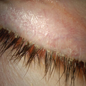
Dr. Rapuano highlighted EBMD, which can cause wrinkles on the surface of the cornea. If it’s mild and in the periphery, it won’t change the corneal shape or curvature, and you can leave it alone, he said. If it’s more significant and in the central cornea, it can create distortions in the vision. These distortions would likely show up on topography or K readings. Patients may notice a little shadow vision, he added.

Source (all): Christopher Rapuano, MD
Salzmann’s nodules, creamy white nodules on the corneal surface, are another issue that Dr. Rapuano discussed. These tend to distort corneal curvature. Often, they are in the periphery, not causing too many visual problems, but they can be an issue if they are closer to the center of the cornea. They can cause astigmatism and potentially irregular astigmatism, he added.
“For all patients we treat, we need to be attentive to the cornea before doing biometry measurements because problems on the surface of the cornea are going to lead to undesirable outcomes,” said John Hovanesian, MD, adding that patient expectations are higher than ever.
The most common issue is dry eye, he said, adding that about three-fourths of those who have it are not symptomatic. “If we’ve got an aim of giving the patient the refractive result they want, we need to pay attention first to dry eye,” he said.
When pterygium and Salzmann’s nodules are visually impactful, they need to be addressed prior to surgery, Dr. Hovanesian said. He avoids putting a toric lens in a patient with a Salzmann’s nodule causing astigmatism because the nodule will cause irregular astigmatism so the correction with the toric is less than ideal. “If you remove the nodule and make the cornea more regular, there may not even be astigmatism, and you’ll at least reduce irregularity,” he explained.
Terry Kim, MD, spoke about many of the previously mentioned conditions as well. He said they may appear benign in nature, but surgeons need to do their due diligence in terms of examining patients, looking for these conditions, and determining if they are functionally or visually significant. “I think that’s a critical step because they can not only affect the visual outcome after surgery, but they can affect biometry measurements.” He cited a paper he co-authored1 that showed that biometry measurements and IOL calculations were significantly impacted in patients with epithelial basement membrane dystrophy and Salzmann’s nodules, which would have resulted in major alterations in spherical and toric IOL power if not treated prior to cataract surgery.

Source: Christopher Rapuano, MD
Are patients already aware of these conditions?
EBMD and Salzmann’s nodules are usually a new diagnosis, Dr. Schallhorn said. “The patients are most commonly unaware of these conditions, as they usually don’t cause symptoms, unless the patient has had previous erosions in the case of EBMD,” she said. “Therefore, it is important to do a careful exam to look for evidence of these before surgery.” She added that patients are usually also unaware if they have mild keratoconus.
Dr. Hovanesian said patients will often attribute poor vision to the cataract rather than corneal problems. “As important as making the diagnosis is the conversation with the patient,” he said. For example, if the patient has dry eye, he will explain to the patient that he or she has two diseases, not one. The first (the cataract) can be fixed, but the second (the dry eye) is up to the patient to treat, sometimes long term. “The emphasis is placed on the patient, and the conversation covers the idea that dry eye will affect vision after surgery,” he said. “I can’t give them perfect vision because their eye is not perfect even with the cataract removed.”
Tests to perform
Corneal topography has become an important part of preoperative evaluation of patients for Dr. Hovanesian. He also gets an OCT of the macula, which is helpful in identifying any macular disease.
Since many dry eye patients are asymptomatic, Dr. Hovanesian said a corneal topography is critical. “If we see corneal staining, that’s not just dry eye, that’s moderate to severe dry eye because the dryness is so advanced that it’s caused a breakdown in the surface,” he said.
Fluorescein staining, Dr. Hovanesian said, can also help show irregularities on the cornea.
Dr. Kim also discussed the importance of using fluorescein, but he warned against using too much, which can flood the tear film, making EBMD hard to spot.
Dr. Kim likes to dab the palpebral conjunctiva with a fluorescein strip moistened with proparacaine and have the patient blink, distributing a thin film that often will highlight the presence, extent, and location of EBMD.
Salzmann’s nodules can be missed, Dr. Kim said, because they often sit in the superior cornea. The physician needs to lift the upper lid to examine the superior cornea to spot these lesions, which can cause a lot of irregular astigmatism.
Dr. Kim also stressed the importance of using corneal topography to see how much a pterygium is pulling on the cornea and causing astigmatism. The pterygium may look minor at first, he said, but the topography can help you determine the extent of central corneal involvement and if it needs to be treated prior to cataract surgery.
“You don’t need a bunch of fancy tests in your office,” Dr. Kim said. Fluorescein staining, tear breakup time, and pressing on the lower lid to look for MGD are all helpful tests that can be easily performed at the slit lamp in 15–20 seconds.
For pterygium, EBMD, and Salzmann’s nodules, doing a good slit lamp exam, lifting the upper eyelid to make sure you visualize the entire cornea, and performing corneal topography are all important steps, Dr. Kim said.
Dr. Rapuano also uses negative staining, which he said can be a valuable tool, particularly for identifying basement membrane dystrophy. These alterations in the tear film indicate elevations in the corneal surface, which are often from subtle EBMD changes. While frequently mild, they can distort the vision and affect biometry, he said.
Most of his cataract patients will also get a topography. He mentioned that this can be helpful to show if a pterygium needs to be treated. People often know if they’ve had a little pterygium for years and might not be bothered, he said. But if the topography is being affected by the pterygium, it should be treated first.
Dr. Schallhorn performs topography on every patient having cataract surgery, calling it incredibly useful for toric planning in patients with normal exams and to detect any irregular corneal contours that might be due to one of the conditions mentioned.
“Topography is a critical part of the preoperative evaluation for any cataract patient,” she said. “This should be combined with a careful exam to identify conditions that could result in a suboptimal outcome and to guide treatment.”
Treatment and when to delay cataract surgery
While Dr. Schallhorn said that no condition is a true contraindication to cataract surgery, addressing issues that can cause problems with biometry is mandatory before proceeding. Patients with dry eye should receive treatment until they have a smooth, stable epithelial surface and are asymptomatic.
Dr. Schallhorn said that patients with EBMD, Salzmann’s nodules, or pterygia causing corneal cylinder should undergo treatment before surgery. Cataract surgery should be delayed to allow for complete epithelial healing, she said, which usually takes 3 months for full stabilization and sometimes longer in patients with EBMD.
Fuchs patients should be counseled about their increased risk for corneal edema. Patients with confluent guttata, morning blur symptoms, or stromal edema should be seen by a corneal specialist because they might benefit from a combined endothelial keratoplasty procedure.
Dr. Hovanesian agreed that it’s important to treat these conditions prior to cataract surgery. For dry eye patients, he said that a month may be an appropriate period to get meaningful improvement. He added that artificial tears, steroid supplements, and other treatments could help with quicker rehabilitation.
When using a surgical treatment for pterygium or Salzmann’s nodules, Dr. Hovanesian noted that most corneal specialists wait 2 months or more postop before proceeding with a cataract procedure. “When you’re altering the corneal stroma, there’s more remodeling and epithelial healing that occurs that may take a couple months,” he said.
Dr. Rapuano described his treatment prior to cataract surgery for both EBMD and Salzmann’s nodules. Typically, you can treat basement membrane dystrophy with epithelial debridement, which he usually combines with diamond burr polishing to make sure all the microscopic basement membrane elevations are gone. This can be done at the slit lamp, Dr. Rapuano added, and it usually takes about 6–8 weeks before the epithelium is smooth for accurate K readings.
For Salzmann’s nodules, he said lamellar keratectomy can be used to treat at the slit lamp. However, he noted the Salzmann’s nodules tend to recur. To try to prevent recurrence, Dr. Rapuano will use mitomycin-C at the time of removal.
Dr. Kim described the criteria used in his study that indicated if a patient’s corneal condition should be treated before cataract surgery: 1) If the patient has central corneal involvement of EBMD or Salzmann’s nodules (central 3–4 mm); 2) Distortion on keratometry or inconsistency on biometry; 3) Irregular astigmatism or “dropout” on corneal topography; 4) Subjective complaints of blurred vision or image ghosting.
Dr. Kim said he and his colleagues treat a lot of these patients with superficial keratectomy and scraping the corneal epithelium. For Salzmann’s nodules, treatment simply involves peeling the lesions with a 0.12 forceps. “For EBMD, we generally perform a superficial keratectomy with a Maloney spatula followed by a small excimer laser PTK procedure,” he said. After both procedures, topical mitomycin-C 0.02% is applied to the treated corneal surface with a circular Merocel sponge for 30 seconds to help prevent recurrence and scar formation.
Dr. Kim said patients are brought back a month after the treatment to confirm complete healing, and sometimes the cataract surgery needs to be delayed. It’s important, he said, to ensure that patients know about these conditions before proceeding with cataract surgery so that they can make an informed decision about treatment with the goal of providing the best visual outcome.
About the physicians
John Hovanesian, MD
Harvard Eye Associates
Laguna Hills, California
Terry Kim, MD
Chief of the Cornea and Refractive Surgery Service
Duke University
Durham, North Carolina
Christopher Rapuano, MD
Chief of the Cornea Service
Wills Eye Hospital
Philadelphia, Pennsylvania
Julie Schallhorn, MD
Residency Program
Associate Director
University of California
San Francisco
San Francisco, California
Reference
- Goerlitz-Jessen MF, et al. Impact of epithelial basement membrane dystrophy and Salzmann nodular degeneration on biometry measurements. J Cataract Refract Surg. 2019; 45:1119–1123.
Relevant disclosures
Hovanesian: Sun Pharma, Novartis
Kim: None
Rapuano: None
Schallhorn: None
Contact
Hovanesian: jhovanesian@harvardeye.com
Kim: terry.kim@duke.edu
Rapuano: cjrapuano@willseye.org
Schallhorn: jschallhorn@gmail.com