Cornea: Back to basics
September 2022
by Liz Hillman
Editorial Co-Director
Corneal imaging for IOL planning is vital, but what instruments to use and what the measurements from these different instruments inform varies. EyeWorld spoke with Douglas Koch, MD, and Kevin M. Miller, MD, to get their thoughts on corneal imaging as part of the preop planning process.
What do you use?
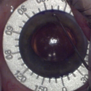
Every new patient in Dr. Koch’s practice gets a Galilei (Ziemer) image. Dr. Koch said this device, which features Placido topography and dual Scheimpflug tomography, gives him the opportunity to assess the anterior surface using the Placido mires. The tomography contributes corneal thickness data and if there is any evidence of ectasia. From there, if cataract surgery is scheduled, Dr. Koch said patients are prescribed an at-home treatment regimen of preservative-free tears, warm compresses, and lid scrubs before coming back for the preoperative visit with biometry readings.
On the day of preop testing, two biometry readings are obtained, an IOLMaster 700 (Carl Zeiss Meditec) and a Lenstar (Haag-Streit). Dr. Koch said both of these contribute valuable corneal data.
“We’re using other devices as well, but those are our mainstays. Patients get three different measurements but on two separate occasions,” Dr. Koch said. “You don’t have to use three different devices, but it’s nice to get at least two measurements, preferably with two different instruments and on separate days, so you get a sense if there are any ocular surface issues that are a concern.”
Dr. Miller also uses a number of different instruments. When selecting the spherical power for a lens, his primary imaging modality is a standard biometer (the Lenstar LS900 or IOLMaster). If he’s doing astigmatism management, he also uses a topographer or tomographer. He has a Galilei but said his workhorse instrument is the Pentacam AXL Wave (Oculus).
Dr. Koch said, in general, if you’re doing presbyopia-correcting IOLs, a tomography measurement should ideally be obtained to confirm a regular cornea that will provide the optical quality needed by the IOL. It also helps the surgeon know if a future enhancement could be possible.

Considerations for quality images and analysis
Dr. Miller said ophthalmologists are often reliant on technicians to ensure quality images and measurements are obtained. The surgeon then analyzes the data, seeing if it makes sense before using it in IOL planning.
1. Head positioning. “If the patient is too low or too high to the measurement axis or too far to the left or right, the topographer or tomographer is going to be centering its measurements on a different part of the cornea rather than the actual center of the cornea. You want the patient to be perfectly centered,” he said.
Tilt of the patient’s head should also be assessed. Even a 5- or 10-degree head tilt greatly affects measurement accuracy, Dr. Miller said.
“Good technicians and good ophthalmologists will make sure the patient is perfectly centered vertically and horizontally, and they will also make sure they are aligned and not tilted,” he said.
Even if the patient is positioned well on the X, Y, and Z axis, Dr. Miller said the tech needs to make sure they are looking in the right direction. He said it’s easy for patients to get distracted and look away, even if the instrument has a target for them to focus on. If patients are not looking at the target, Dr. Miller said it can result in measurements showing astigmatism or coma that does not exist.

2. Flag abnormalities and reimage, if necessary. Surface abnormalities (especially dry eye), contact lenses, cosmetic material, or debris in the tear film can “trip up measurements,” Dr. Miller said. If the measurements or imaging appears off, Dr. Miller said that the hope is that you will notice before the patient goes home so they can be reassessed.
“I obtain all the imaging before they see me. If I see issues, I can send them back before they leave,” he said.
Dr. Koch said to look for consistency (and inconsistency). “If you see inconsistency between two devices with regard to overall corneal power or astigmatic values, that’s a red flag that requires further investigation, and further measurements are needed, perhaps after more intensive treatment of the ocular surface to make the best recommendation and best choice for the patient,” he said.
Dr. Koch said technicians at his practices are trained to look at the ease with which measurements can be obtained and any warning signals coming from the devices, for example, distortions in the mires produced from the Galilei.
3. Know when reimaging won’t improve results. Dr. Miller added that some corneas are irregular—those with Salzmann’s nodules or map-dot-fingerprint, for example—and will not improve with repeat imaging. The surgeon needs to know when repeat imaging won’t make a difference unless prior intervention is taken.
4. Get multiple measurements. Dr. Koch reiterated that his practice gets multiple measurements on different occasions, which allows him to look for consistency and inconsistencies. While he said that most surgeons aren’t getting two different measurements on two different occasions, he thinks it’s a best practice.
“Surgeons who are doing refractive cataract surgery by and large are doing two different measurements, but I think it’s not common to do two different measurements and different measurements on different days,” he said.
5. Look at the total cornea. If you have access to a device that looks at both anterior and posterior surfaces, Dr. Miller thinks you should consider the total cornea in your IOL planning and not just the anterior surface.
“I think a lot of people still use a biometer, an IOLMaster or Lenstar, to do their astigmatism planning,” Dr. Miller said. “The problem with those devices is they only see a small portion of the anterior surface of the cornea; they don’t see the total cornea. You have to make assumptions about the back surface of the cornea if you’re only measuring the anterior surface of the cornea. You can use the Barrett formula for that, for instance.”
Dr. Miller noted that some, like Warren Hill, MD, have put forth the idea that the posterior surface measurements might not be all that accurate and measuring the anterior surface and making assumptions might produce an equally adequate result. Dr. Miller said he doesn’t share this perspective but some physicians do.

6. Look for LASIK and PRK. Dr. Miller said that 10–15% of his cataract practice includes patients who have had prior PRK or LASIK. “On any given day, there is a good chance that I am operating on someone who has had prior laser vision correction surgery,” he said.
The catch is some of them don’t include these procedures in their medical history. Even if you ask them if they’ve had prior eye surgery, many say no. Dr. Miller said they don’t necessarily consider LASIK to be “eye surgery.” Topography, tomography, or OCT “will save you from doing surgery on people you didn’t know had refractive surgery,” he said.
“You think you should be able to see this prior surgery when you examine them, but you can’t see PRK, and in some eyes it is difficult to see LASIK. The edge of the LASIK flap is just so well healed, it looks normal,” he said.
A biometry measurement is unlikely to reveal prior refractive surgery, Dr. Miller said, unless the cornea is significantly flatter than you think it should be, but even that is nuanced with lower treatments.
7. Onboarding new equipment. When you bring in a new imaging modality, Dr. Miller said to take time to evaluate it against the imaging you were using before.
“I will continue doing the old thing, image them with the new thing, and retrospectively look at if I chose to go with the old thing, will the new thing give me a better outcome?” he said. “I think every time we bring in new equipment, you need to evaluate it in some way and … not just suddenly flip over to the new and abandon what you were doing before and maybe getting good results with.”

Source (all): Kevin M. Miller, MD
‘Garbage in, garbage out’
Every instrument has its own quirks, and you’ve got to pay attention to various factors to get a quality image and measurement for IOL selection, Dr. Miller said.
[template id=14580]“There is a famous computer saying: Garbage in, garbage out. If you have a person not looking in the right direction, their eyes are dry, their head is tilted 30 degrees, guess what? You’re going to get terrible information by which to operate on,” he said.
Lens power formulas are looking for inputs, values, Dr. Miller continued. There are many possible inputs, but the most important ones are corneal curvature and axial length. Of these, he said axial length is the most important to nail, with small errors in measurement resulting in large errors in lens power. Biometers do a good job of this, Dr. Miller said.
For corneal power, Dr. Miller said most ophthalmologists still use biometers, but he thinks Scheimpflug and OCT devices that look at the front and back surface of the cornea are best for calculating a toric lens.
“I think measuring corneal power with these devices and planning out your astigmatism management is better than using a simple biometer,” he said.
About the physicians
Douglas Koch, MD
Department of Ophthalmology
Cullen Eye Institute
Baylor College of Medicine
Houston, Texas
Kevin M. Miller, MD
Kolokotrones Chair in Ophthalmology
David Geffen School of Medicine at UCLA
Los Angeles, California
Relevant disclosures
Koch: Alcon, Bausch + Lomb, Carl Zeiss Meditec, Johnson & Johnson Vision
Miller: Alcon, Oculus
Contact
Koch: dkoch@bcm.edu
Miller: kmiller@g.ucla.edu