ASCRS News
April 2022
Non-diffractive wavefront shaping extended depth of focus (EDOF) intraocular lens: visual performance and patient-reported outcomes
Thomas Kohnen, MD, Kerstin Petermann, MSc, Myriam Böhm, MD, Eva Hemkeppler, Wasim Ahmad, Lisa Hinzelmann, Katarzyna Pawlowicz, MD, Tyll Jandewerth, MD, Christoph Lwowski, MD
A prospective, single-arm study at the Department of Ophthalmology at Goethe University in Frankfurt, Germany, evaluated visual performance and patient-reported outcomes after bilateral implantation of the Vivity IOL (Alcon), a non-diffractive extended depth of focus IOL. Sixteen patients (32 eyes) were included in the study, with a target of emmetropia for both eyes. At 3 months postop, mean spherical equivalent was –0.16±0.37 D. Binocular UDVA at distance (4 m), intermediate (80 cm, 66 cm), and near (40 cm) was 0.01±0.05 logMAR, 0.05±0.05 logMAR, 0.07±0.06 logMAR, and 0.25±0.11 logMAR, respectively. Some optical phenomena were noted, but 88% of these patients said they would choose the same lens again. Sixty-three percent of patients didn’t have any optical phenomena. Photopic and mesopic contrast sensitivity was good.
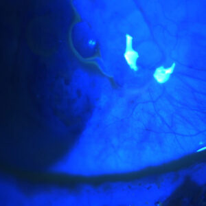
Clinical and histopathological findings in the dead bag syndrome
Catherine Culp, MD, Phillip Qu, MD, Jason Jones, MD, Nicole Fram, MD, Gregory Ogawa, MD, Samuel Masket, MD, Nick Mamalis, MD, Liliana Werner, MD, PhD
Clinical and histopathological analysis of capsular bags with suspected “dead bag syndrome” was conducted to provide a better understanding of why some capsular bags become diaphanous and floppy, unable to support an IOL, years after surgery. The study was conducted at the John A. Moran Eye Center in Salt Lake City, Utah, and included 10 cases of suspected dead bag syndrome. Seven explanted capsular bags underwent histopathological examination and five explanted IOLs were examined microscopically. The authors noted capsular thinning and/or splitting in the capsular bags. Two capsular bags had no lens epithelial cells and five had only a few lens epithelial cells on the inner surface of the capsule. The IOLs were three-piece silicone lenses or single-piece hydrophobic acrylic lenses; four of these optics were “unremarkable,” but one had some granular pigment deposition. The authors concluded that the syndrome appears to have an absence of secondary proliferation of lens epithelial cells and fibrotic changes, as well as degradation of the capsule. They advocated for further studies to better understand the etiology of the syndrome.
Real world incidence of monofocal toric IOL repositioning: analysis of the American Academy of Ophthalmology IRIS Registry
Brent Kramer, MD, John Berdahl, MD, Xiaolin Gu, MD, Mohinder Merchea, OD
Repositioning of TECNIS toric IOLs (Johnson & Johnson Vision) were five times higher than AcrySof toric IOLs (Alcon), according to a retrospective study that used data from the American Academy of Ophthalmology IRIS Registry. Eyes that had cataract surgery with one of these two lenses, implanted in 2016 and 2017, were included in the study. The rate of reoperation for realignment within a year of implantation was evaluated. There were 2,013 eyes that received a TECNIS toric within the study period and 4,469 that received an AcrySof toric. The incidence of repositioning within the first year was 3.1% in eyes that had a TECNIS implant compared to 0.6% in eyes that had an AcrySof.