Cornea
September 2022
by Clara Chan, MD
Cornea Editor

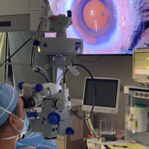
One of my retina colleagues recently referred a phakic diabetic patient to our cornea clinic who 3 weeks after her first intravitreal anti-VEGF injection developed a red eye, photophobia, and tearing. On slit lamp exam, she had a 1 x 2 mm fluffy endothelial plaque surrounded by a ring of satellite lesions about 4.5 mm in diameter and a 1 mm hypopyon. There was no epithelial defect, no history of contact lens wear, no contact with plant matter, no history of topical steroid use, and no travel to tropical climates. Without an epithelial defect and the findings being in the posterior cornea, we were hesitant to culture. It was suspected that the infection was somehow seeded during the anterior chamber tap done after intravitreal injections to avoid intraocular pressure elevations. Based on the clinical appearance, oral voriconazole (which has excellent intraocular penetration) and compounded amphotericin 0.15% drops every hour were prescribed.

Source: Clara Chan, MD
Fungal infections in Canada are fairly rare due to our cooler climate, but over the last decade, our tertiary center is gradually seeing more.1 When approached with a potentially complex clinical presentation, one must go back to the basics of obtaining a good history and physical exam. One of the things I love about ophthalmology, and cornea in particular, is that we are able to efficiently make visual diagnoses. During our years in training and in practice, we see such high volumes that each of us has created an image database in our memories to hone our pattern recognition abilities.
I hope you enjoy this issue of EyeWorld, where we have articles that review the diagnosis and management of fungal keratitis; that break down the differences between evaporative dry eye and aqueous tear deficient dry eye, which is important for tailoring patient treatments; and that detail how to optimize your corneal imaging modalities to ensure excellent images and accurate analysis.
Reference
- Trinh T, et al. Clinical and microbiological analysis of fungal keratitis in Toronto, Canada: A 20-year study. Med Mycol. 2022;60:myac047.