Cover Feature: MIGS roundup
July 2017
by Liz Hillman
EyeWorld Staff Writer
“Hybrid” MIGS device launches in U.S. market
Options—that’s what many physicians say the XEN45 Gel Stent (Allergan, Dublin, Ireland), one of the latest implants to join the market of surgical glaucoma procedures, gives them.
Joseph Panarelli, MD, assistant professor, Department of Ophthalmology, New York Eye and Ear Infirmary of Mount Sinai, New York, said he thinks the XEN has broad applications in a variety of patients.
Nathan Radcliffe, MD, clinical assistant professor, Department of Ophthalmology, New York University Langone Medical Center, New York, emphasized that XEN can be a standalone procedure or coupled with cataract surgery. It also leaves the option open for more aggressive glaucoma surgery, if needed, later down the line.
Arsham Sheybani, MD, assistant professor of ophthalmology and visual sciences, Washington University School of Medicine, St. Louis, discussed the XEN as a “hybrid” between microinvasive glaucoma surgery (MIGS) and traditional glaucoma surgery.
“I say hybrid for two reasons. I think it is a hybrid between traditional surgery and MIGS because it’s minimally invasive, it’s very controlled, but it also accesses the subconjunctival space route, which right now is our best way to lower pressure with a trab or a tube. It’s not quite MIGS; it’s not quite the higher risk glaucoma surgeries,” Dr. Sheybani said. “The other reason I call it a hybrid is it’s almost a hybrid of a trabeculectomy and a tube. It’s a tube itself, but the flow starts immediately in the [subconjunctival] sub-Tenon’s space, and we have to use mitomycin just like we do with our trabeculectomies.”

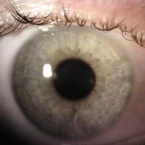
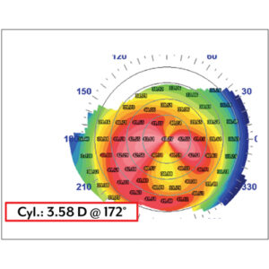
Source: Joseph Panarelli, MD
About the implant
The XEN received approval from the U.S. Food and Drug Administration (FDA) in November 2016 and launched in the U.S. in early 2017.
The translimbal implant targeting the subconjunctival space for ab interno bleb formation consists of a 6-mm porcine gel material crosslinked with glutaraldehyde, 45 µm in diameter, that swells and becomes flexible when implanted.
“The advantages there are it will accommodate to the tissue and this should limit the erosion,” Dr. Radcliffe explained. “If you had something rigid under the conjunctiva, erosion would be more common, and the swelling is important because once it has been placed, it’s not likely to move and fixes itself into position.”
The composition of the material itself also does not elicit a fibrotic response, Dr. Sheybani said. The procedure is creating a controlled wound in the eye, he explained, and the body naturally wants to cause fibrosis. Certain materials naturally elicit a fibrotic response, so having a material that doesn’t is a theoretical advantage, Dr. Sheybani said.
Patient selection
Per on-label uses, the XEN is indicated for refractory glaucoma patients with failed previous surgical treatment and for patients with primary open-angle glaucoma (POAG) or those with pseudoexfoliative or pigmentary glaucoma with open angles who are not responsive to the maximum medical therapy.
However, some doctors consider off-label applications as well. Dr. Panarelli said he would consider it in ocular hypertension patients who are intolerant of medical therapy as well as those with mild to moderate glaucoma. Dr. Sheybani said, “For me, it’s that the surgery is so predictable that I prefer it for even earlier intervention because those are the eyes where you would hate to take someone with good vision and minimal field loss but high pressures, you would hate to subject them to the aggressive surgery … but in this case, we have an operation that seems to have less risk.” Dr. Sheybani said he would use it in angle closure and pediatric cases, which are also considered off-label.
Dr. Radcliffe has used the XEN in a number of different scenarios, but his favorite candidate is a young, phakic individual with excellent vision.
“A 24-year-old patient of mine had juvenile primary open-angle glaucoma, and his pressures were 40 mm Hg on all of the medications. I did XEN Gel Stents in both eyes, and he’s now off all of his drops with pressures of 8,” Dr. Radcliffe said. “That’s an ideal scenario, but now he’s going to have a life with almost no hardware in his eye and with good, controlled pressures, and that’s what he needed because he’s so young. I point out this case because this is his first glaucoma surgery after laser trabeculoplasty, so I think this is a first-line approach for patients, and it should be because of its safety profile.”
Dr. Sheybani said this procedure is suited for patients with the least chance of scarring. He also said a little bit of proptosis is helpful in early cases because it can be difficult to access a deeply set eye. Avoid the XEN all together in those eyes with scarring in the area where the stent would be placed.
Dr. Panarelli said he would avoid placing the stent in patients with advanced disease, finding it difficult to achieve low IOPs in those cases. He acknowledged, however, there are some patients with more advanced disease who might insist on avoiding trabeculectomy or tube shunt surgery; in these cases, he would be willing to try the XEN as it has a more reasonable chance of obtaining a low pressure when compared to other MIGS procedures.
Surgical technique
Implanting the XEN is a completely new procedure to everyone who does it, according to Dr. Sheybani.
“It’s not that the technique is easy, but the procedure is reproducible and low risk compared to traditional glaucoma surgeries.”
Arsham Sheybani, MD
“It’s not a surgery that any one surgeon has an advantage for doing. By that I mean whether you are a glaucoma, comprehensive, or cornea [specialist], none of us has an advantage over another because it’s so different in how it’s done,” he explained. “The nice thing is that the risks of intraop complications are so low when you look at studies across the board. It’s not that the technique is easy, but the procedure is reproducible and low risk compared to traditional glaucoma surgeries.”
According to Allergan’s website, the implant is injected ab interno through a clear corneal incision with a pre-loaded, single-use injector with a 27-gauge needle. It is positioned with about 2 mm in the subconjunctival space, 3 mm left intrasclerally, and 1 mm in the anterior chamber. Dr. Sheybani said one doesn’t need to be so dogmatic when it comes to these measurements. “You just want enough of a tunnel to where it’s going to remain in there and enough poking under the conjunctiva and enough poking into the [anterior chamber]. … You want to make sure the two lumens are open and not blocked by tissue,” Dr. Sheybani said, likening the stent to a straw.
He recommended using whatever type of anesthesia you prefer and just making sure patients are well anesthetized before starting the case, as movement on their part could compromise stent placement.
Before inserting the stent, Dr. Radcliffe uses 40 micrograms—0.2 cc of the 0.2 mg/ml—of mitomycin-C, injecting it very posteriorly and massaging it forward.
Paul Palmberg, MD, PhD, professor of ophthalmology, Bascom Palmer Eye Institute, Miami, explained that the use of mitomycin-C (MMC) is needed to retard the formation of additional resistance to aqueous outflow in Tenon’s capsule or the conjunctiva.
“Injecting the MMC allows the procedure to be done in a ‘suture- free’ manner as no conjunctival incision needs to be made,” Dr. Panarelli said.
If the XEN is not combined with cataract surgery, Dr. Sheybani said the clear corneal incision should be angled toward the targeted quadrant of stent placement, with the surgeon making sure the injector sits in that position well without hitting the cheekbone or speculum. A second instrument through a paracentesis helps stabilize the inserter as the tip of the needle is poked through the sclera in the eye, which Dr. Sheybani said should be firm with a cohesive viscoelastic. Dr. Sheybani prefers to place the stent well in the subconjunctival space, which can look like you might poke through the conjunctiva, he acknowledged. If you’re worried about that, he recommended creating a small bleb with balanced salt solution or viscoelastic to tent it up. With forward pressure on the needle, making sure it doesn’t move up or down or side to side, Dr. Sheybani presses the slider until it feels like it won’t go any further.
“Then you relax your hands very slowly, equilibrate the system, and slowly withdraw the injector out of the eye,” leaving the stent in place, Dr. Sheybani said.
Dr. Panarelli does not use fluorouracil routinely postop, preferring to keep his patients on more potent steroid medications like difluprednate. Dr. Radcliffe said digital ocular compression can be used to help XEN flow.
“If I have a XEN that’s working partially in a patient and [pressure] is starting to creep up, I’ll encourage the patient to compress the eye and to even massage the area over the bleb itself in order to encourage flow,” he said.
Overall, Dr. Radcliffe said that while XEN implantation does take some finesse, he found the learning curve to be rapid. “Getting the stent in the perfect position away from the cornea in the angle but not near the iris just takes a little intuition and can be achieved in the first few cases,” he said.
Complications
While Dr. Sheybani said intraoperative complications are low, postoperative complications could arise if the stent is not placed properly.
If too much of the stent is left in the anterior chamber, repositioning is required, but this becomes a challenge as the implant is hydrated and not easily advanced, Dr. Panarelli said. When more than 2 mm of the stent is left beneath the conjunctiva, there is a greater possibility that it could become kinked and/or project upward if caught in Tenon’s, he added.
If reimplantation is needed, the procedure becomes more difficult due to reduced visibility (bleeding from the first pass attempt) and increased patient discomfort, Dr. Panarelli said, adding that there’s also the likelihood that more needle track passes will induce more scarring at the implantation site.
When the XEN is coupled with cataract surgery, if the stent is placed immediately after lens removal, the eye is softer and can make stent placement more difficult, Dr. Panarelli said. It’s unknown yet if the inflammation associated with cataract surgery will impact long-term stent function, he added.
As for needling, Dr. Radcliffe said he needles about a third of his cases but thinks this is because he’s trying to get patients to very low pressures without the use of medication.
“If I was more likely to tolerate a pressure of 17 on one drop, then I would be less likely to needle patients,” he said. “But because I’m trying to get patients to have pressures of 10 off drops, I am needling, and I’m having great efficacy.”
Dr. Palmberg explained that the dimensions of the XEN were calculated to produce a pressure gradient from the anterior chamber to the subconjunctival space of 8 mm Hg at normal aqueous flow, using the Hagen-Poiseuille equation. This greatly reduces the risk of persistent hypotony compared to trabeculectomy. He said that the XEN and the InnFocus MicroShunt (Santen, Osaka, Japan) are the only minimally invasive procedures that have produced low to normal average IOPs—11 mm Hg for the InnFocus MicroShunt at 3 years1 and 12.6 mm Hg for the XEN at 2 years.2
Dr. Palmberg cited a recent reevaluation of the Collaborative Initial Glaucoma Treatment Study that suggests achieving pressures in the low teens has an advantage for even modestly damaged POAG patients, based on an average 10% recovery of the visual field and the increased safety of surgical options.3 The XEN and InnFocus MicroShunt potentially offer safer surgical options to reach such pressures, and the MicroShunt is currently being compared to MMC trabeculectomy in ongoing FDA and European trials.
Dr. Panarelli thinks the best time to needle is when pressures first begin to creep up. “Early needling to disrupt scar tissue formation is easily accomplished as there is still flow through the device, and this can be done in the office,” he said, adding that he reserves more aggressive needling for the operating room, especially when the stent becomes “capped,” resulting in a flat-appearing bleb.
Another complication could be bleb leaks—though Dr. Panarelli called them uncommon—which should only result if there is perforation of the conjunctiva. Though he has yet to encounter it, Dr. Panarelli said his biggest concern is erosion, which would more likely occur when the device fails and comes in close contact with the eyelid. He hasn’t had any cases of early or late onset hypotony, but Allergan does mention this as a possibility. The small luminal diameter of the stent provides sufficient resistance to mostly avoid these issues, he said.
The XEN, like other MIGS procedures, leaves open the possibility of trabeculectomy and tube shunts, if needed, by the patient later. Because the XEN bleb tends to be more nasal, Dr. Radcliffe said a trabeculectomy could still be done at 12:00, and as many tube shunts as needed could be performed.
Dr. Panarelli said reimbursement has been a challenge thus far. While it’s likely to change in the future, he has found that prior authorization is required by many insurance plans, or the patient pays out of pocket. Dr. Radcliffe also said that while he’s found some insurance plans cover the XEN, it’s important to have a policy where patients understand they will pay out of pocket and be reimbursed by insurance, if covered.
“I don’t hesitate to ask patients to pay out of pocket, reminding them that if the insurance reimburses, they’ll be reimbursed. But this is the case where if patients pay several thousand dollars for a stent, they’re going to see excellent intraocular pressures, they’re going to have their glaucoma problems resolved, they’re going to be very thankful and happy that they made this investment in their vision,” Dr. Radcliffe said, adding that he’s optimistic about the future of XEN insurance reimbursement moving forward.
Dr. Sheybani doesn’t implant the XEN without prior insurance approval, not wanting patients to pay out of pocket when there are other options covered by insurance.
“These operations may replace trabeculectomy and tube-reservoir shunts in primary glaucoma surgery if the clinical trials show equal efficacy and greater safety. They will likely replace MIGS because of greater effectiveness and comparable safety, and will conceivably compete with medical therapy as initial treatment for glaucoma,” Dr. Palmberg said.
References
- Batlle JF, et al. Three-year follow-up of a novel aqueous humor microshunt. J Glaucoma. 2016;25:e58–65.
- Ahmed IK, et al. Use of a 45 μm ab-interno subconjunctival gel-stent with adjunctive mitomycin-C for the treatment of uncontrolled open angle glaucoma. 2015 AGS Annual Meeting.
- Musch DC, et al. Visual field improvement in the collaborative initial glaucoma treatment study. Am J Ophthalmol. 2014;158:96–104.
Editors’ note
Drs. Panarelli, Radcliffe, and Sheybani have financial interests with Allergan. Dr. Palmberg has financial interests with Allergan and Santen.
Contact information
Palmberg: ppalmberg@med.miami.edu
Panarelli: jpanarelli@NYEE.EDU
Radcliffe: drradcliffe@gmail.com
Sheybani: sheybaniar@wustl.edu