Cornea: Cornea editor’s corner of the world
March 2012
by Faith A. Hayden
EyeWorld Staff Writer

The term dendrite is defined as a branching treelike figure. This term describes a shape. When clinicians see a “branching treelike figure” in the corneal epithelium, the most immediate thought is herpes simplex virus (HSV) keratitis. It is important for clinicians to realize that a dendrite (referring to the shape) is not always the infectious epithelial lesion of HSV. There are many other “dendritic” lesions of the corneal epithelium that are not due to HSV and have been referred to as “pseudodendrites.”
It is very common to see patients with healing epithelial defects or neurotrophic epitheliopathy present with a “dendrite” or a branching epithelial lesion. These patients are often placed on topical antivirals, which leads to delayed epithelial healing and persistence of this pseudodendrite. In addition, the label of HSV is often permanently attached to the patient.
A detailed history, careful slit lamp examination, and close monitoring of the clinical course will assist the clinician in determining the correct diagnosis and treatment for these challenging patients. Clara Chan, M.D., and Mark Mannis, M.D., give us their insights for the examination of the patient with a “dendrite.”
—Edward J. Holland, M.D., cornea editor
Many epithelial lesions have a dendritic shape. Although most of these lesions are pseudodendrites, they are frequently misdiagnosed as herpes simplex virus (HSV). The shape differences between these dendrites are subtle, but do exist. EyeWorld spoke to two experts on the subject who weighed in on the telltale signs.
True HSV dendrites
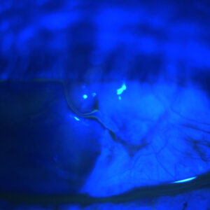
Authentic HSV epithelial disease typically presents as a unilateral, branching lesion with little end bulbs, which stain very brightly with fluorescein.
“It’s usually in the context of previous herpetic infections,” said Mark J. Mannis, M.D., professor and chair, ophthalmology and vision science department, University of California, Davis. “It can present as a keratitis. It can be very large, it can be very tiny, and therein lies the difficulty. It can present in someone who doesn’t have a history of diagnosed herpes simplex.”


Source (all): Clara C. Chan, M.D.
True HSV also progresses in stages, said Clara C. Chan, M.D., professor of ophthalmology, University of Toronto. Vesicles will form and appear as small, raised lesions at first and will not stain with fluorescein.
“Vesicles should not be confused with punctate epithelial erosions, which stain positively with fluorescein,” Dr. Chan said. “Within approximately 24 hours, these vesicles coalesce to form the classic dendrite that most ophthalmologists recognize.”
“The virus bounces from cell to cell, so each lesion develops one cell at a time, which is why you get this linear lesion because there are cells falling out and virus cells filling in,” Dr. Mannis explained.
HSV, however, doesn’t always present this way. For example, immune-compromised patients may have vesicles only and no dendrite, said Dr. Chan. On the other hand, patients with recurrent HSV keratitis and intact immune systems may have these vesicles early on in a recurrence prior to examination.
If a patient has an enlarged dendrite that’s not linear, it’s a geographic ulcer. A classic clinical observation is a scalloped-shaped boarder, not to be confused with healing abrasions and epithelial defects from neurotrophic keratopathy, which have smooth borders.
“Many clinicians associate prior topical steroid use with geographic ulcers, but a geographic ulcer from HSV may form without prior topical steroid use as well,” Dr. Chan said.
Pinpointing pseudodendrites
“The term pseudodendrite is a little bit of a misnomer,” Dr. Mannis said. “We call them pseudodendrites because they are not herpes simplex, but they are in fact dendrites.”
Varicella zoster (VZV) dendrites are quite different from HSV. For example, unlike HSV, which stains brightly, VZV stains minimally. It has a “stuck on appearance” that’s elevated and presents as “convoluted clusters,” Dr. Mannis said. VZV can take on a medusa-like form, with its many branches and lack of end bulbs.


Source: Edward J. Holland, M.D.
Furthermore, VZV is typically found in the periphery of the cornea, Dr. Chan said. They’re also more superficial and have no central ulceration.
Pseudodendrites have a wide variety of causes including neurotrophic epitheliopathy, Acanthamoeba, healing epithelial defect, and recurrent erosion syndrome, Dr. Chan said.
In neurotrophic epitheliopathy, the pseudodendrite is often in the interpalpebral region of the cornea. “The patient has already undergone treatment with either oral or topical antivirals,” Dr. Chan explained. “Suspect if the infectious keratitis lasts more than 2 weeks.”
Acanthamoeba may be to blame if a patient has a history of improper contact lens wear and hygiene, and uses contacts while swimming or in hot tubs.
“Symptoms can range from foreign body sensation to pain out of proportion to the clinical appearance,” Dr. Chan said. “Isolated epithelial pseudodendrites can appear as granular, cystic-appearing epithelium with little or no stromal inflammation.”
If a patient has a history of corneal abrasion, no prior history of herpes simplex, and no ulceration upon exam, the pseudodendrite is likely due to a healing epithelial defect. However, if the patient’s affected eye was recently cut or injured, consider recurrent erosion syndrome.
“[Patients have] classic clinical history of awaking at night or upon first awakening sudden pain, ‘like my eye lid is ripping the skin off my eye,'” Dr. Chan explained. “Always check the fellow eye to determine if there are any signs of epithelial basement membrane degeneration.”
In rare cases, pseudodendrites can be caused by tyrosinemia type 2. Those patients will have associated findings of mental retardation and hyperkeratosis of the hands and feet.
Reference
Holland EJ and Schwartz GS. Classification of herpes simplex virus keratitis. Cornea 1999; 18:144-154.
Editors’ note
The doctors mentioned have no financial interests related to this article.
Contact information
Chan: clara.chan@gmail.com
Mannis: mjmannis@ucdavis.edu